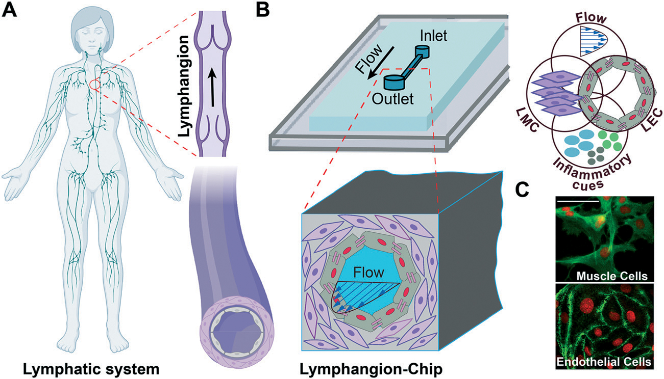
Fig. 1.

Lymphangion-chip: microfluidic model of a lymphangion. (A) Illustration of the human lymphatic vessel consisting a lymphangion which is the unit between the two adjacent valves and (B) an engineering drawing of the lymphangion-chip with co-cultures of lymphatic endothelial cells (LECs) and lymphatic muscle cells (LMCs) that is leveraged to analyze the responses to flow and inflammatory cues. (C) A representative confocal image set of on-chip LMCs (green: F-actin) and LECs (green: VE-cadherin) under co-culture conditions. Scale bar: 50 μm.
