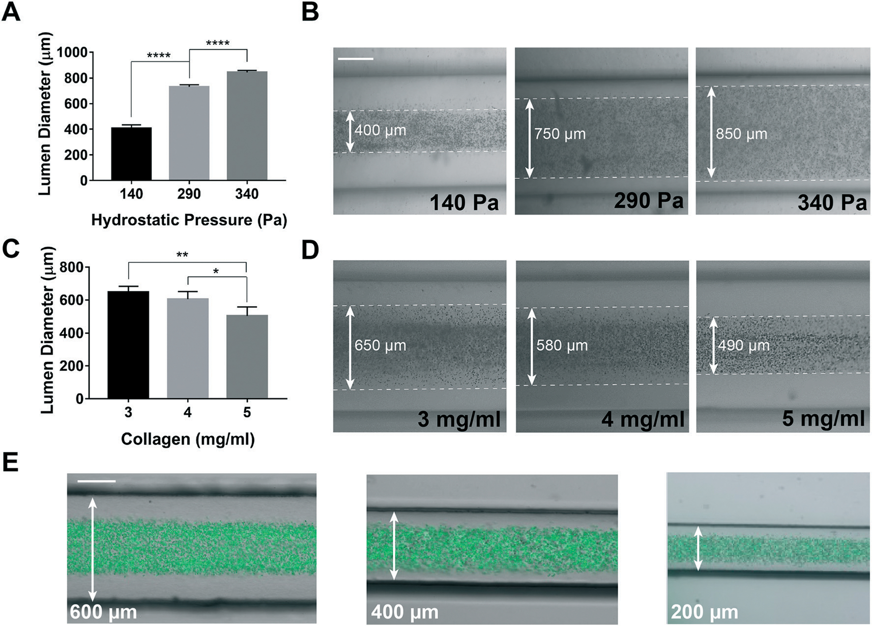
Fig. 3.

Engineering and tuning of the lymphangion-chip. (A) Graph and (B) representative images of engineering the lumen inner diameter and the muscle tissue thickness by setting the hydrostatic pressure and (C and D) collagen concentration. (E) Engineering the outer vessel diameter by running the GLP method in chips with various channel widths (200–600 μm) (mixture of green fluorescent beads with cell medium specifies the lumen). All scale bars: 200 μm; *p < 0.05, **p < 0.005, ****p < 0.0001; n = 3–5 for all the experiments.
