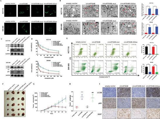
Figure 5.

CircATG4B‐222aa but not circATG4B increases the autophagy and induces oxaliplatin in CRC cells in vitro and in vivo. A) Puncta‐like staining detection of HCT116 and SW480 transfected with four vectors. Scale bars, 10 µm. B) Transmission electron microscopy analysis of autophagy of HCT116 and SW480 transfected with four vectors. Scale bars, 2 µm. C) LC3B‐II/LC3B‐I proportion of HCT116 and SW480 transfected with four vectors. D) CCK‐8 detection of cell viability by oxaliplatin in HCT116 and SW480 transfected with four vectors. E) Flow cytometry analysis of cell apoptosis by oxaliplatin in HCT116 and SW480 transfected with four vectors. F) Nude mice xenografts were formed by HCT116 cells transfected with empty‐vector, circATG4B, circATG4B‐mut, and circATG4B‐222aa, respectively. Then, sixteen days after injection, oxaliplatin was injected intraperitoneally. G) Tumor volumes were monitored during the time course. H) Tumor weights were also measured at the end of this study. I) IHC analysis of LC3B, p62, and Ki67 in tumor nodules. Scale bars, 20 µm. **p < 0.01, ***p < 0.001; ***p < 0.001 empty vector versus circATG4B group, ### p < 0.001 circATG4B‐mut versus circATG4B‐222aa group (H).
