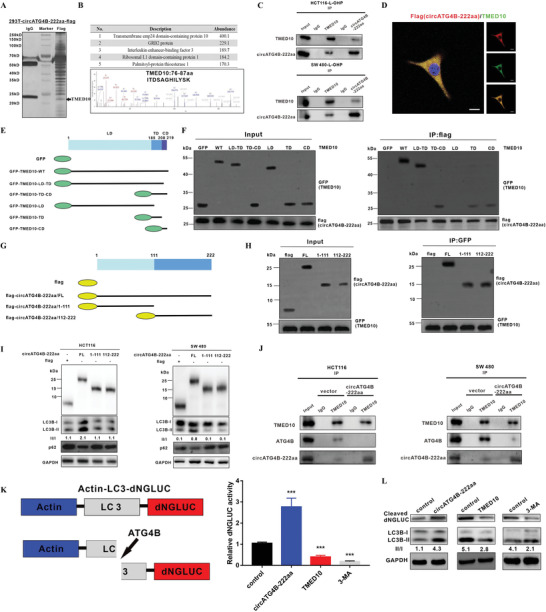
Figure 6.

CircATG4B‐222aa protects against the effect of ATG4B induced by TMED10‐mediated inhibition. A) Total proteins from flag‐circATG4B‐222aa plasmid‐transfected HEK‐293T cells were extracted. The proteins coimmunoprecipitated with antibody against flag were separated via SDS‐PAGE. B) Upper panel, list of the top five differentially expressed proteins identified by mass spectrometry. Lower panel, TMED10 was identified by LC/LC‐MS. C) The interaction of TMED10 and circATG4B‐222aa was detected by immunoprecipitation in CRC cells. D) Flag‐tagged circATG4B‐222aa was transfected into HCT116 cells and immunofluorescence was performed using anti‐flag and anti‐TMED10 antibody. Scale bars, 10 µm. E) Schematic of domain structure of TMED10 and GFP‐tagged TMED10 mutants. F) HEK‐293T cells were transfected with flag‐tagged circATG4B‐222aa and GFP‐tagged full‐length or TMED10 fragments, followed by IP using anti‐flag antibody. G) Schematic diagrams showed the wild‐type circATG4B‐222aa and its truncation mutants. H) HEK‐293T cells were transfected with GFP‐tagged TMED10 and flag‐tagged circATG4B‐222aa mutants, followed by IP with anti‐GFP antibody. I) Western Blotting assays showed the levels of autophagy in cells after transfection of wild‐type circATG4B‐222aa ORF or truncation mutants. J) After overexpression of circATG4B‐222aa, the binding capacity between TMED10 and ATG4B was monitored by Co‐IP. K) Left panel, schematic diagram of the quantification of ATG4B activity using an assay based on a luciferase‐release system. Right panel, HCT116 cells transfected with pEAK12‐Actin‐LC3‐dNGLUC were transfected with control, or circATG4B‐222aa overexpression or TMED10 overexpression, or treated with 3‐MA. The supernatants were collected, and the relative luciferase activity was measured. L) The collected supernatants were analyzed by Western blotting using an anti‐luciferase and anti‐LC3 antibody.
