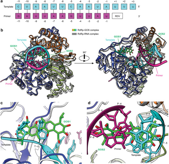
Figure 4.

Structural comparisons of the GOS‐bound and RNA‐bound RdRp complexes. a) Definition of the base position of RTP and primer‐template RNA. b) Overall views of the SARS‐CoV‐2 RdRp–GOS complex overlapped with the RNA‐bond SARS‐CoV‐2 RdRp structure (PDB ID 7BV2). The RNA‐bond SARS‐CoV‐2 RdRp structure is shown in gray, the template RNA is cyan, and the primer RNA is purple. c) Zoom in view of GOS1 overlapped with RNA template strand from the RNA‐bond SARS‐CoV‐2 RdRp structure. d) Zoom in view of GOS2 overlapped with the RNA template and primer strands from the RNA‐bond SARS‐CoV‐2 RdRp structure.
