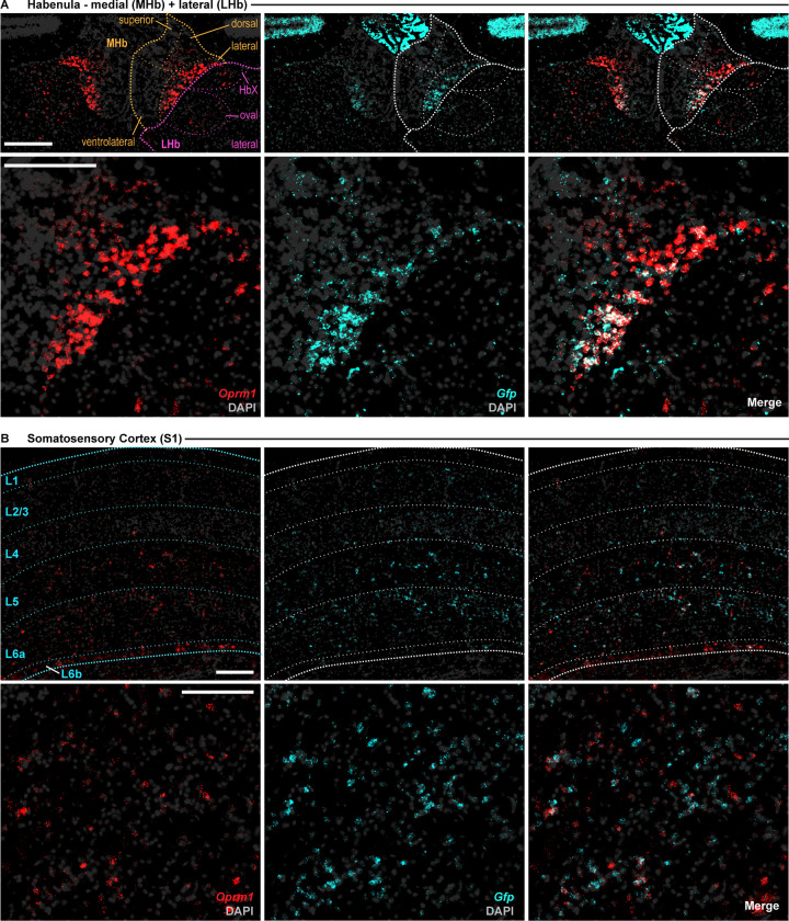
Fig 3. Fluorescence in situ hybridization of Oprm1Cre; RosaLSL-GFP-L10a mice confirms selective reporter expression in Oprm1+ cells in mouse habenula and cortex.
Overlap of Oprm1 (red) and GFP (Cyan) mRNA in (A) the medial and lateral habenula; and (B) Primary Somatosensory Cortex. Left images: Oprm1 signal (red) and DAPI (grey); Middle: GFP signal (cyan) + DAPI (grey); Right images: Merge of all signals. Top images: Entire ROI with cortical layers and/or subnuclei indicated; bottom images: large image of area of densest Oprm1 signal. L1-6b: indicates cortical layer; HbX: HbX subdivision of the habenula; MHb, medial habenula; LHb, lateral habenula; S1; primary somatosensory cortex. Signal was detected using the RNAScope Multiplex Fluorescence V2 kit. 16μm slices were imaged using the BZ-X800 Viewer software in conjunction with a BZ-X Series automated Keyence microscope. Images were taken at 40x magnification. Mice (n = 2) were homozygous for both transgenes. All scale bars: 200μm.

