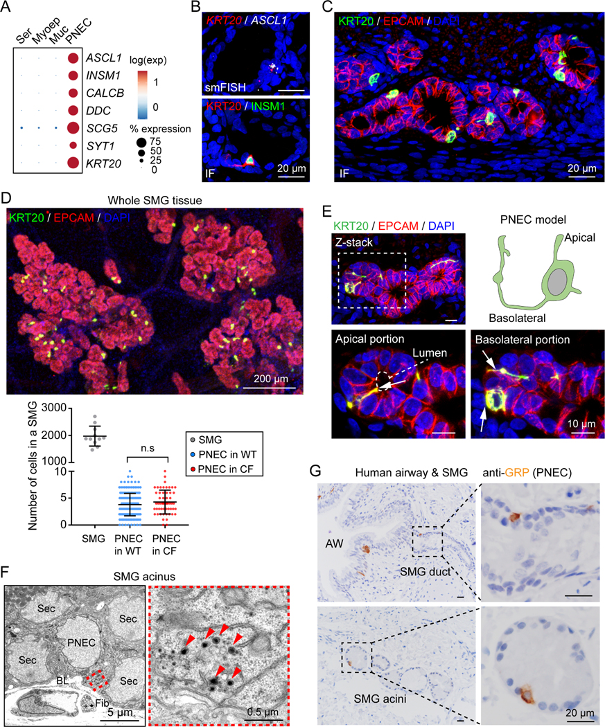
Figure 1 |. PNECs are localized in porcine and human SMGs.
A) PNEC marker gene expression in single-cell RNA sequencing (scRNA-seq) data from porcine SMGs (Yu et al., 2022). Ser, serous cell; Myoep, myoepithelial cell; Muc, mucous cell.
B, C) Single-molecule fluorescence in situ hybridization (smFISH) and immunofluorescence (IF) of PNEC markers (ASCL1, INSM1, KRT20) in SMGs (EPCAM+).
D) Quantification of PNECs in individual SMGs. Upper panel, immunostaining of PNECs (KRT20+, green) in whole mount SMGs (EPCAM+, red). Lower panel, summary of total cells and PNECs in individual SMGs. Each data point represents number of cells or PNECs in a SMG. P value was calculated between WT and CF based on individual SMGs; p > 0.05 is considered as not significant (n.s.), unpaired Student’s t test; n = 3–4 pigs.
E) SMG PNEC reconstruction. Enlarged images show apical and basolateral portions (arrows) of a PNEC connecting to the acinar lumen (dashed outline) and other SMG cells.
F) Representative transmission electron microscopic images of rare cells (PNECs) with dense core vesicles (arrowheads) in SMGs. Sec, secretory cell of SMGs; BL, Basal lamina; Fib, fibroblast.
G) Immunohistochemistry of gastrin-releasing peptide (GRP, human PNEC marker) in human airways (AW) and SMGs.

