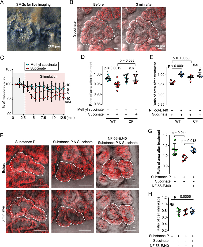
Figure 4 |. Extracellular succinate stimulates SUCNR1-dependent SMG contraction.
A) Representative reflected light image of SMGs in newborn pig trachea after cartilage and smooth muscle layers were removed.
B) Representative images of SMGs (EPCAM+, red) before and after succinate treatment. Closed lines outline the selected SMG regions for area measurement.
C) Summary of SMG area treated with succinate or methyl succinate during live imaging of SMGs. 3 glands each group.
D, E) Quantification of succinate-induced SMG contraction. Relative SMG area 3 minutes after a treatment. Panel D, SMGs were treated with 10 mM methyl succinate or succinate. Panel E, Airway surface epithelia were removed. SMGs were pre-incubated with or without SUCNR1 antagonist NF-56-EJ40 (5 μM) and induced with succinate. Data points are the mean of 3 SMGs from each pig; p values are shown in graph; p > 0.05 is considered as not significant (n.s), Student’s t test.
F, G) Succinate prevents SMG swelling due to mucus secretion triggered by substance P. Panel F, representative images of SMGs that were pre-incubated with or without NF-56-EJ40 and treated with substance P and/or succinate. Panel G, quantification of SMG swelling response to substance P and/or succinate treatment. P values are shown in graph, n = 5 pigs, paired Student’s t test.
H) Quantification of acinar cell shrinkage under succinate and/or substance P stimulation. P = 0.0006, n = 5 pigs, one way ANOVA. See also Figure S5.

