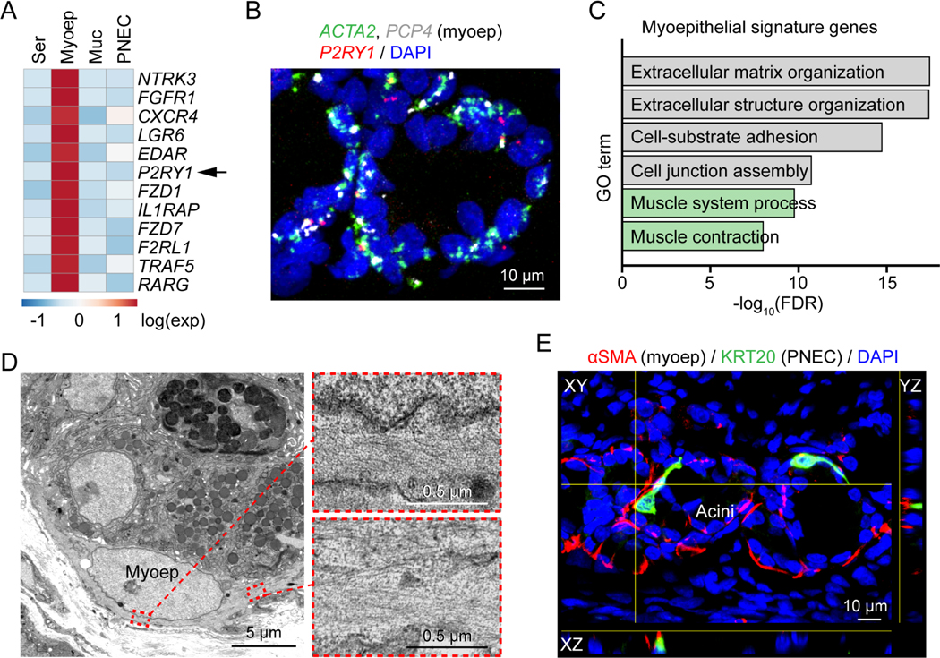
Figure 6 |. SMG myoepithelial cells express the purinergic receptor P2RY1.
A) Heatmap of myoepithelial signature receptors in SMG scRNA-seq data (Yu et al., 2022). Genes are ranked by false discovery rate from the lowest to the highest. Ser, serous cell; Myoep, myoepithelial cell; Muc, mucous cell.
B) Representative smFISH image of ACTA2, P2RY1, and PCP4 in SMGs. Myoep, myoepithelial cell.
C) Gene ontology (GO) analysis of myoepithelial-cell signature genes. Muscle contraction related pathways are highlighted in green. FDR, false discovery rate.
D) Representative transmission electron microscopy images of myoepithelial cells in SMGs. Enlarged images show microfilaments of the myoepithelial cell.
E) Representative image of PNEC (green) and myoepithelial cell (myoep, red) proximity in SMGs.

