Figure 3.
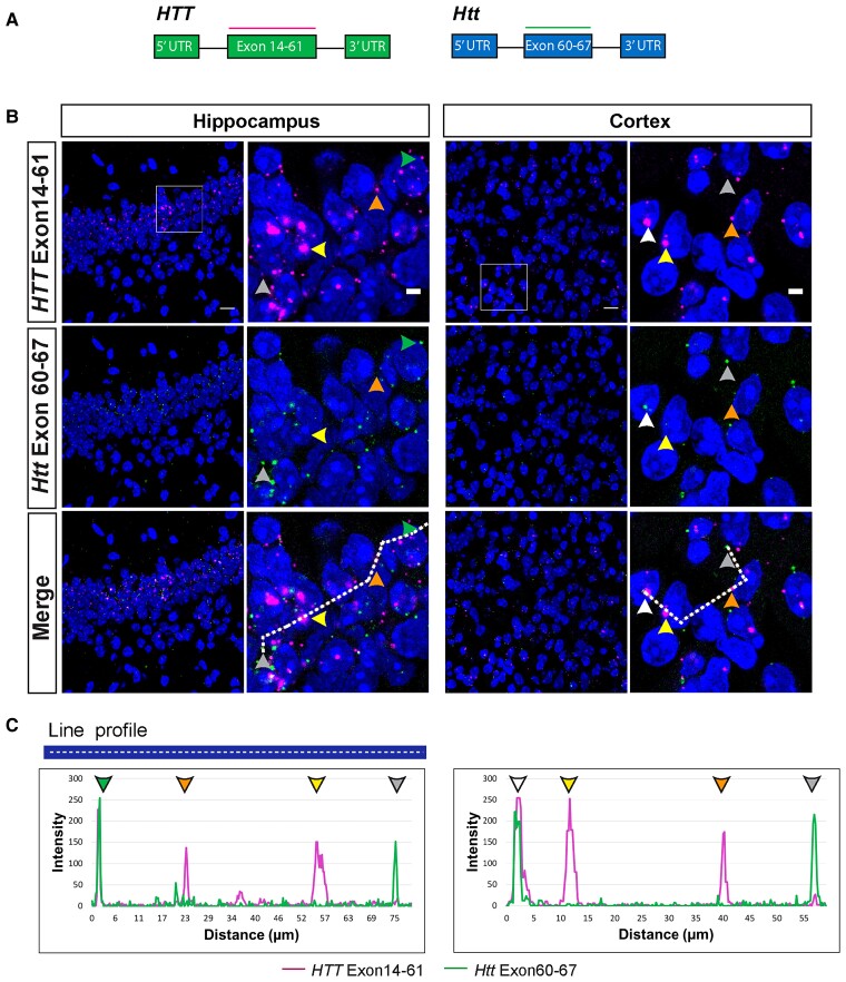
Mouse Htt transcripts were present in the nucleus and cytoplasm, whereas human HTT mRNAs were retained in nuclei in YAC128 brains. (A) Schematic location of the full-length human (exons 14–61) and mouse (exons 60–67) huntingtin RNAscope probes. (B) Full-length human HTT (magenta) was present in large nuclear clusters (yellow arrowheads) and as single transcripts in the extranuclear space (orange arrowheads). Full-length mouse Htt (green) was predominantly detected outside of the nucleus (grey arrowheads). Human and mouse huntingtin transcripts only rarely co-localized in both the nucleus (white arrowheads) and cytoplasm (green arrowheads). (C) Intensity profiles of full-length mouse and human huntingtin transcript signals. Peak intensities were recorded along the white dashed line shown in the merged image in panel (B). Yellow arrowheads indicate large nuclear clusters containing human HTT, orange arrowheads indicate human HTT transcripts outside of the nucleus, grey arrowheads indicate mouse Htt transcripts outside of the nucleus, white arrowheads indicate co-localization of human HTT and mouse Htt inside the nucleus and green arrowheads indicate co-localization of human HTT and mouse Htt in the cytoplasm. A statistical analysis of the transcript subcellular location is presented in Supplementary Fig. 1C and D. Nuclei were stained with DAPI (blue). The wild-type control sections from mice at 2 months of age are shown in Supplementary Fig 3A and B. YAC128 (n = 4). Scale bar = 20 μm in the main image and 5 μm is the cropped magnified image.

