Figure 2.
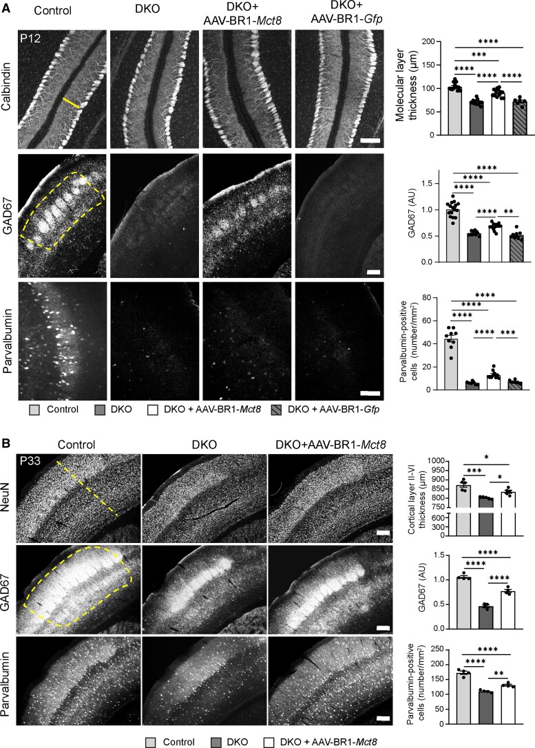
AAV-BR1-Mct8 treatment improves neuronal morphology and gene expression. (A) Administration of AAV-BR1-Mct8 at P0 to DKO mice increased thickness of the molecular layer in the cerebellar vermis as well as the relative fluorescence intensity of GAD67- and parvalbumin-positive interneurons of the somatosensory cortex at P12. Administration of AAV-BR1-Gfp at P0 to DKO mice had no effect. One-way ANOVA: molecular layer thickness, F(3/40) = 50.25, P < 0.0001; Holm–Sidak’s post hoc test. Welch’s ANOVA: GAD67, W(3/21.43) = 58.74, P < 0.0001; parvalbumin-positive interneurons, W(3/15.37) = 64.72, P < 0.0001; Tamhane's T2 post hoc test. Scale bar = 100 µm (top and bottom panel); Scale bar = 200 µm (middle panel). (B) Administration of AAV-BR1-Mct8 at P0 improved the thickness of the somatosensory cortex layers II–VI, the relative fluorescence signal intensity of GAD67 and the number of parvalbumin-positive interneurons in the cortex at P33. One-way ANOVA: cortical thickness, F(2/12) = 16.4, P = 0.0004; GAD67, F(2/9) = 102.0, P < 0.0001; parvalbumin-positive interneurons, F(2/9) = 63.01, P < 0.0001; Holm–Sidak’s post hoc test. Scale bar = 200 µm. Each dot represents one animal. Means ± SEM are shown. Dashed line indicates region of interest. *P < 0.05; **P < 0.01; ***P < 0.001; ****P < 0.0001.

