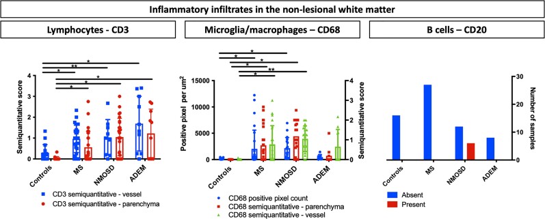
Figure 2.
Inflammation in the non-lesional white matter. Bar graphs show the characteristics of the inflammatory infiltrate in the non-lesional white matter of five controls, five acute disseminated encephalomyelitis (ADEM), 14 multiple sclerosis (MS) and four neuromyelitis optica spectrum disorder (NMOSD) donors. Only samples without demyelination were considered. Left, CD3+ lymphocytic infiltration was increased compared with controls in all the demyelinating CNS diseases with highest values observed in ADEM cases. Middle, There was a significant increase in CD68+ microglial infiltration in MS and NMOSD cases compared with controls. Left, B-cells were typically present in NMOSD cases but were not found in MS and ADEM cases (chi-square 14, P = 0.001). CD3+ lymphocytic and CD68+ microglial infiltration were quantified using a semiquantitative scores for parenchymal and one for perivascular inflammation; automated pixel counting software was also used for CD68. B-cell inflammation was classified as with or without CD20-positive cells in the non-lesional white matter. Bars display the mean values, the whiskers represent CI95%. The asterisks indicate significant post hoc pairwise comparisons (*P < 0.05, **P < 0.001).

