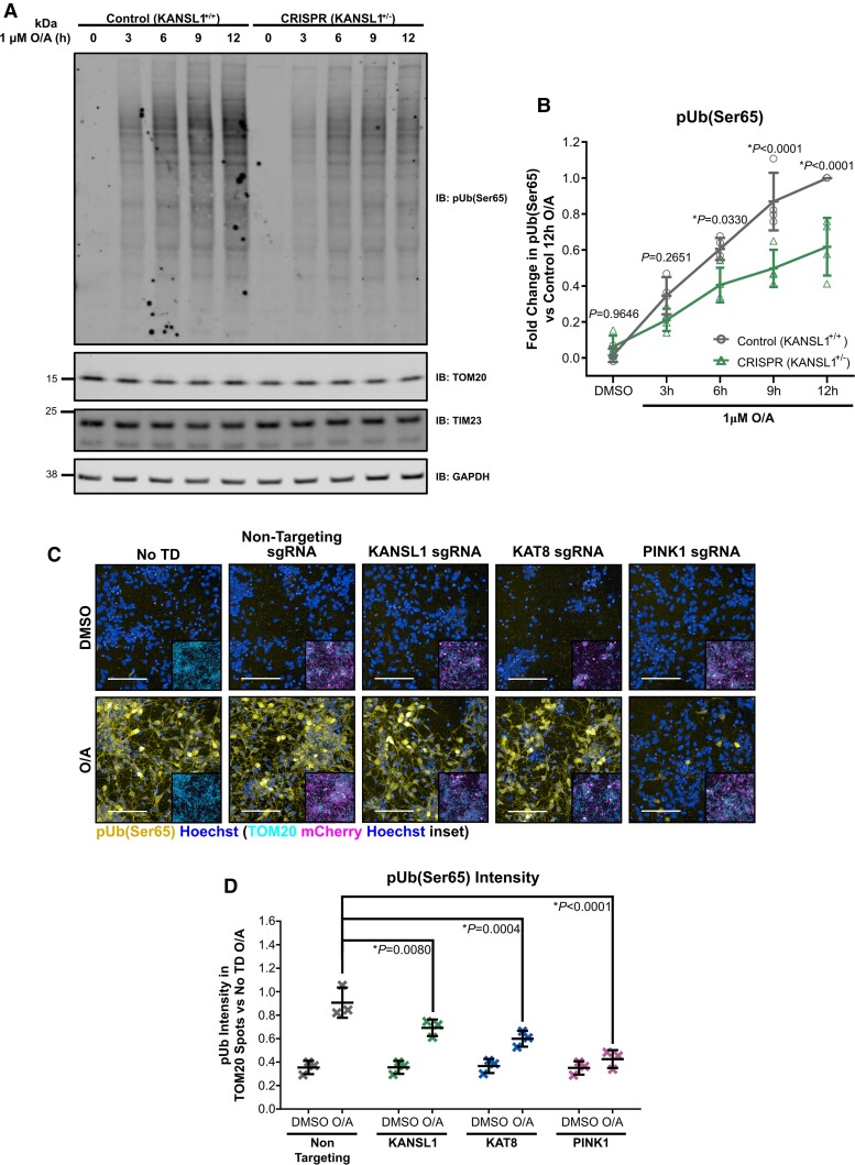
Figure 7.
pUb(Ser65) levels are reduced in isogenic iNeurons with heterozygous KANSL1+/- loss of function and CRISPRi-i3N iNeurons with KANSL1 and KAT8 sgRNA KD. (A) Representative IB of isogenic d17 iNeurons with/without heterozygous LoF frameshift mutation in KANSL1 treated with 1 µM O/A over a 12 h extended time-course. (B) Quantification of pUb(Ser65) in A (n = 4 inductions, two-way ANOVA with Dunnett’s correction). (C) Representative images of pUb(Ser65) (yellow) with Hoechst nuclei counterstain (blue) following treatment of non-transduced (No TD), non-targeting, KANSL1, KAT8 and PINK1 sgRNA KD d17 CRISPRi-i3N iNeurons with 1 µM O/A versus DMSO for 9 h. Inserts show staining for TOM20 (cyan) and mCherry transduction reporter (magenta) with Hoechst nuclei counterstain (blue) for the same field of view. Scale bar = 100 µm. (D) Quantification of pUb(Ser65) intensity in TOM20 defined mitochondrial area in D (n = 3 inductions, two-way ANOVA with Dunnett’s correction). Data are shown as mean ± SD.

