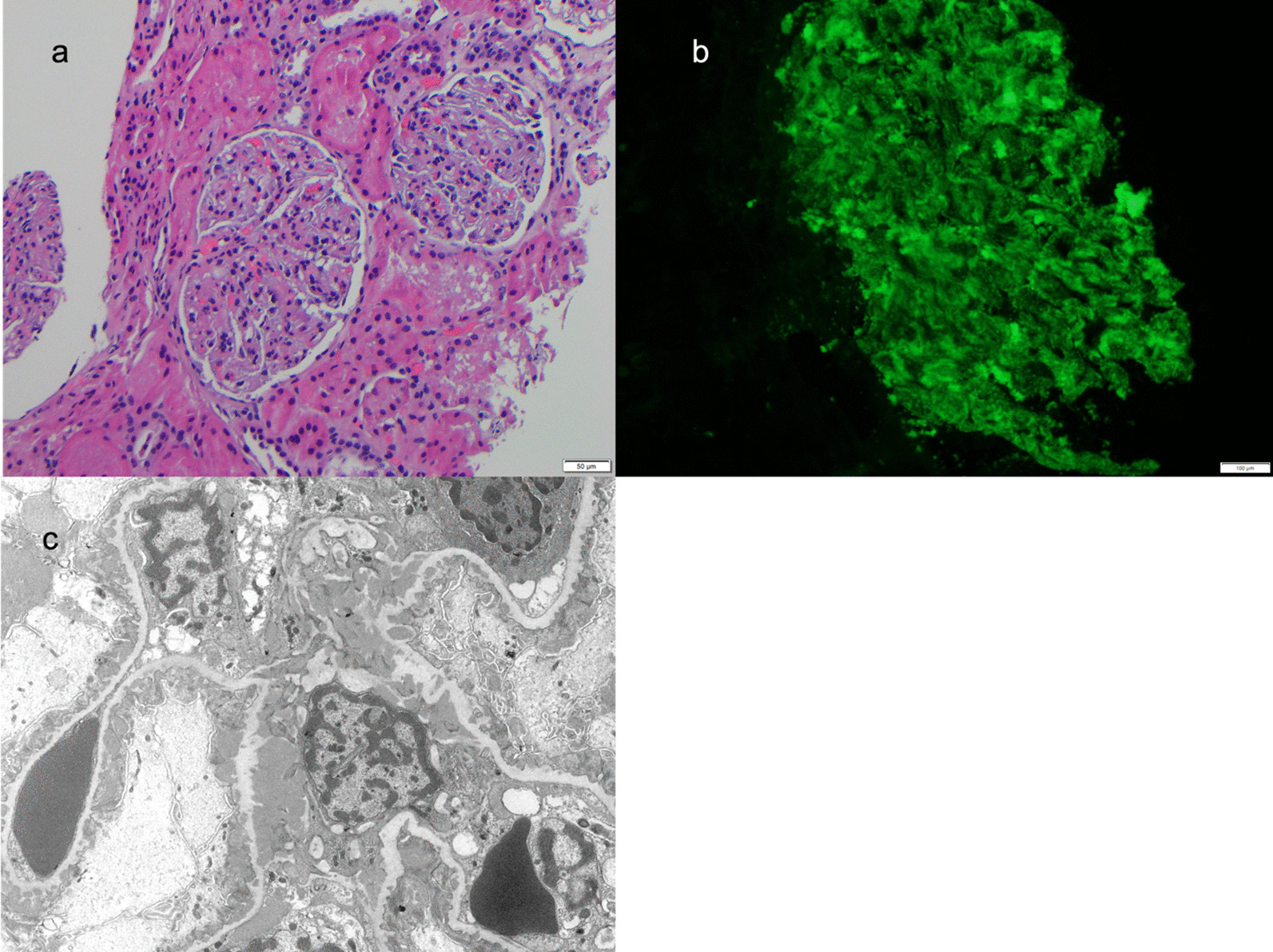
Fig. 1.

Kidney Biopsy showing a light microscopy, b immunofluorescence and c electron microscopy. a By light microscopy, glomeruli had a mild increase in mesangial matrix and cells with intact appearing basement membranes. The interstitium, tubules, and small vessels were normal except for abundant protein droplets the tubular epithelium. b Granular mesangial and peripheral staining was present on direct antibody immunofluorescence with antibodies against IgA, IgG, IgM, C3, and C1q. c Ultrastructural examination confirmed abundant mesangial and paramesangial deposits. Numerous early subepithelial membranous deposits were associated with small basement membrane spikes. Subendothelial tubuloreticular bodies were present (not shown)
