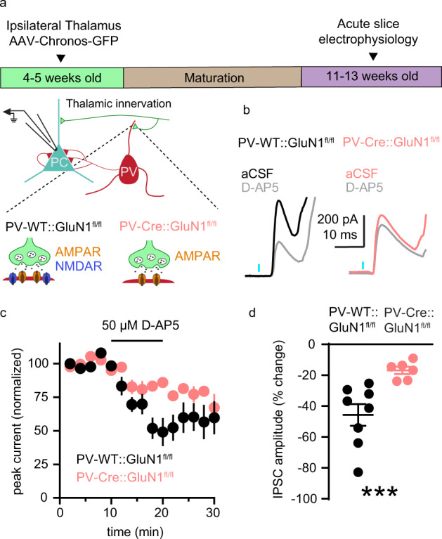
Fig. 6. PV+ interneuron NMDAR contribute to thalamus-evoked FFI in mPFC pyramidal neurons.

a Schematic of the experimental approach used to measure the contribution of PV+ interneuron NMDARs to thalamus-evoked FFI in layer 5/6 pyramidal neurons (0 mV holding potential) in acute coronal slices of adult mPFC. The effect of D-AP5 on IPSC amplitude was compared in mice with GluN1 expression intact (black; PV-WT::GluN1fl/fl) or GluN1 knocked out (pink; PV-Cre::GluN1fl/fl) from PV+ interneurons. b Representative recordings of feedforward IPSCs in slices from PV-WT::GluN1fl/fl (left) or PV-Cre::GluN1fl/fl conditional knockout (right) mice in aCSF (black or pink) and during application of 50 µM D-AP5 (gray). c Average time course of normalized feedforward IPSC amplitude in PV-WT::GluN1fl/fl (black; n = 8) or PV-Cre::GluN1fl/fl conditional knockouts (pink; n = 6) in response to D-AP5 application. d D-AP5 decreases feedforward IPSC amplitude more in PV-WT::GluN1fl/fl (n = 8 neurons; 45.71 ± 7.0%) than in PV-Cre:: GluN1fl/fl conditional knockouts (n = 6 neurons; 16.87 ± 2.37%; U = 0; p < 0.001). Group data (c, d) represented as mean ± SEM. ***p < 0.001; Mann–Whitney test for unpaired comparisons (d).
