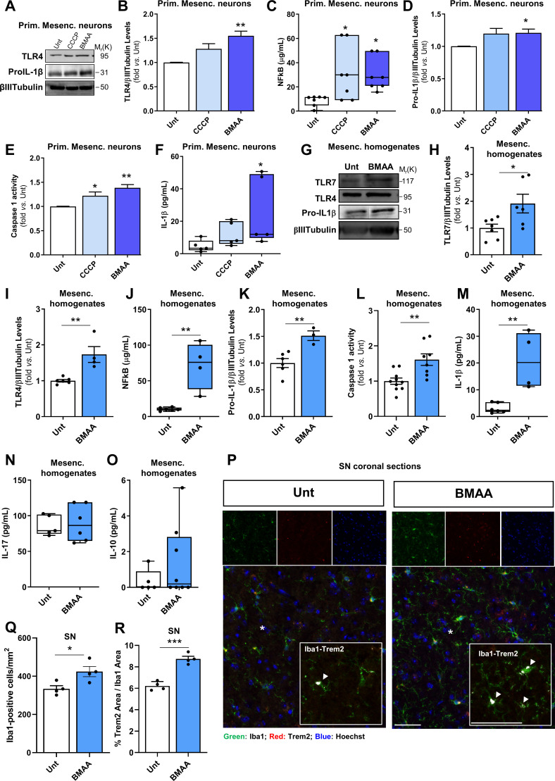
Figure 5.
β-N-methylamino-L-alanine (BMAA) activates brain neuronal innate immunity in vitro and in vivo. Primary mesencephalic neuronal cultures from naive mice were treated with 1 µM CCCP for 2 hours and 3 mM BMAA for 48 hours. (A) Representative immunoblot for toll-like receptor (TLR)4 and pro-interleukin (IL)-1β levels. Blots were re-probed for βIII-Tubulin to confirm equal protein loading. (B) Densitometric analysis of the levels of TLR4 was normalised with βIII-Tubulin (n values for all conditions=4, untreated (Unt) vs CCCP, p=0.076; Unt vs BMAA, **p=0.0023). (C) Nuclear factor kappa-B (NF-κB) levels were calculated using NF-κB p65 ELISA kit. Values are μg/mL (n values for all conditions=7, Unt vs CCCP, *p=0.0039, Unt vs BMAA, *p=0.013). (D) Densitometric analyses of the levels of pro-IL-1β were normalised with βIII-Tubulin (n values for all conditions=5, except BMAA=4, Unt vs CCCP, p=0.1213; Unt vs BMAA, *p=0.044). (E) Caspase-1 activation (n values for all conditions=4, Unt vs CCCP, *p=0.047, Unt vs BMAA, **p=0.002). (F) IL-1β levels in the isolated cytosolic fraction was determined using an IL-1β ELISA kit. Values are pg/mL (n values for all conditions=5, Unt vs CCCP, p=0.5928; Unt vs BMAA, *p=0.018). Homogenates from the mesencephalon of mice treated with or without BMAA were examined. (G) Representative immunoblot for TLR7, TLR4 and pro-IL-1β levels. The blots were re-probed for βIII-tubulin to confirm equal protein loading. (H) Densitometric analyses of TLR7 levels normalised against βIII-tubulin (n values for Unt=7 and BMAA=6, Unt vs BMAA, *p=0.027). (I) Densitometric analyses of TLR4 levels normalised against βIII-tubulin (n values for Unt=6 and BMAA=4, Unt vs BMAA, **p=0.004). (J) NF-κB levels were calculated using NF-κB p65 ELISA kit. Values are μg/mL (n values for Unt=6 and BMAA=4, Unt vs BMAA, **p=0.002). (K) Densitometric analyses of pro-IL-1β levels normalised against βIII-tubulin (n values for Unt=6 and BMAA=3, Unt vs BMAA, **p=0.007). (L) Caspase-1 activation (n values for Unt=10 and BMAA=8, Unt vs BMAA, **p=0.003). (M) IL-1β levels were determined using an IL-1β ELISA kit. Values are pg/mL (n values for Unt=6 and BMAA=4, Unt vs BMAA, **p=0.003). (N) IL-17 levels were determined using an IL-17 ELISA kit. Values are pg/mL (n values for Unt=5 and BMAA=6, Unt vs BMAA, p=0.8029). (O) IL-10 levels were determined using an IL-10 ELISA kit. Values are pg/mL (n values for Unt=5 and BMAA=8, Unt vs BMAA, p=0.483). (P–R), Iba1 and Trem2 expression in SN from Unt and BMAA-treated mice by immunofluorescence. (P) Representative images of brain coronal sections stained with Iba1 (microglial and macrophage-specific calcium-binding protein), Trem2 (triggering receptor expressed on myeloid cells 2) and Hoechst 33 342 as nuclei marker in SN. Enlarged boxes show the area of Trem2 (white pixels) contained in Iba1 signal. (Q) Quantification of the number of Iba1+ cells per mm2 (n values for all conditions=4, Unt vs BMAA, *p=0.028). (R) Percentage of Trem2 area contained in Iba1 expression (n values for all conditions=4, Unt vs BMAA, ***p=0.0002). Scale bars are 50 µm. Data represent mean+SEM. Statistical analysis: One-way analysis of variance followed by Dunnett’s test was performed in B, D–E. Kruskal-Wallis test followed by Dunn’s test was performed in C and F. Unpaired Student’s t-test was performed in H–N and Q–R and Mann-Whitney test in O.

