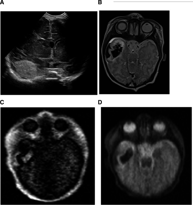
Figure 2.
(A) Five-day-old baby boy with intraparenchymal haemorrhage in the anterior right temporal lobe on an ultrasound examination. (B) The conventional MRI examination demonstrated the intraparenchymal and subpial haemorrhage well on the T2-weighted images. The portable MRIs also showed the extent of the intraparenchymal and subpial haemorrhage in the right temporal lobe on the T1-weighted (C) and T2-weighted images (D).

