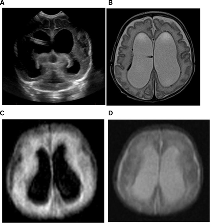
Figure 3.
(A) An 88-day-old baby girl with hydrocephalus with dilated lateral ventricles with an intraventricular catheter in place shown on head ultrasound. (B) The conventional MRI examination showed severe dilation of the bilateral lateral ventricles on the T2-weighted images with hemosiderin staining of the right lateral ventricular lining. This portable MRI also showed similar dilation of the bilateral lateral ventricles on FLAIR (C) and T2-weighted images (D). The hemosiderin staining of the ventricular lining is not imaged well on the portable MRIs. FLAIR, fluid-attenuated inversion recovery.

