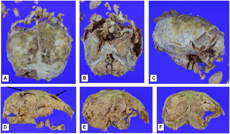
Figure 2.
Macroscopic findings at time of formalin-fixed brain evaluation. Dorsal view of brain covered by adherent calvarial dura mater and epidural tan-brown granular material (A); frontotemporal view of brain partially covered by dura mater (brainstem and cerebellum indistinct) (B); lateral view of brain and dura (side uncertain) (C); coronal section of posterior frontal cerebral hemispheres and adherent calvarial dura mater, showing bilateral red-brown subdural discoloration (arrows), as well as possible dural icterus (green discoloration), and with focally discernible tan-brown cerebral cortex overlying tan white matter, with focal yellow-orange foci (D); coronal sections at parieto-occipital and occipital/cerebellar levels (E, F), with similar changes to those seen in panel (D). (See text for additional description.)

