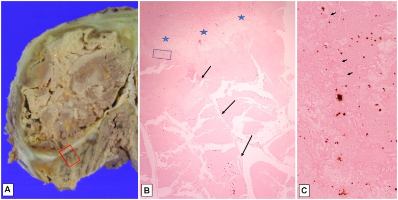
Figure 9.
Gross fibrous adherence of tentorium to overlying occipital cortex and underlying cerebellar cortex, with yellow-orange discoloration (A); histologic section (B, H&E, 20×) of red-boxed area in (A), showing subpial molecular layer adherent to tentorium (delimited by blue stars), and underlying autolyzed cerebellar cortical folia (long arrows) with cholesterol clefts and artifactual cracking of tissue; blue-boxed area in (B), showing “ghost-like” Purkinje cell profiles (short arrows), as well as orange-brown bilirubin crystals (C, H&E, 200×).

