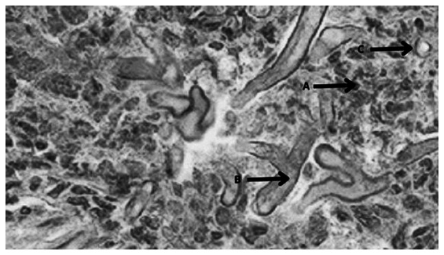
Figure 2.

Histological image of mucormycosis (periodic acid-Schiff stain; magnification, x100). Pro-inflammatory cells are present co-existing with necrotic tissue (arrow A). Septate or pauciseptate fungal hyphae are visible all throughout the connective tissue specimen (arrow B). Sporangiophores containing spores are also seen, suggesting mucormycosis (arrow C).
