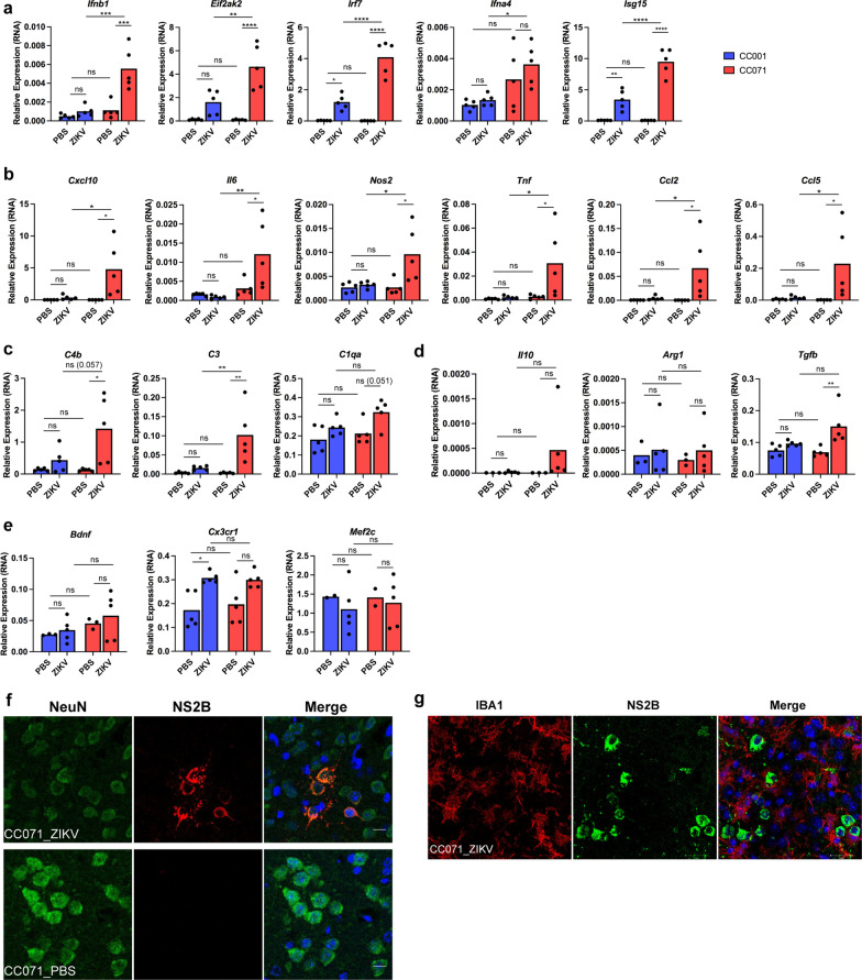
Fig. 4.
ZIKV-infection induces the activation of microglia and of IFNs-I and pro-inflammatory responses in the brain of immunocompetent CC071 mice, in the absence of microglia infection. CC001 and CC071 mice (5–6 week-old) were necropsied at day 6 following IC inoculation of either PBS or 105 FFU of ZIKV. a–e As compared to non-susceptible CC001 mice, ZIKV infection of susceptible CC071 mice induced a significantly higher expression of mRNAs coding a for IFNB and ISGs and for factors associated with the b the pro-inflammatory response and c the complement cascade without significantly affecting genes associated with d the anti-inflammatory response and e the homeostatic “off” state of microglia as determined by RT-qPCR analysis of total brain extracts with respect to Hrpt1 used as reference gene. Symbols represent individual mice. Data from n = 5 CC001 and CC071 mice respectively are means without s.d. with significance assessed by two-way ANOVA Tukey’s multiple comparisons test. P-value < 0.0001 (****), < 0.001 (***), < 0.01 (**), < 0.05 (*) and ns not significant; P-values comprised between 0.05 and 0.1 (considered as near significant) are indicated. f, g ZIKV induced microglia activation in CC071 mice brains while only infecting neurons as determined by immunofluorescence and confocal microscopy with neurons labeled with anti-NeuN (green), microglial cells with anti-Iba1 antibody (red), ZIKV-infected cells with anti-NS2B antibody (red or green) and DNA labeled with DAPI (blue). Single confocal sections (z projection) and the corresponding merge images are shown. Scale bars in f 10 µm and g 20 µm

