Fig. 7.
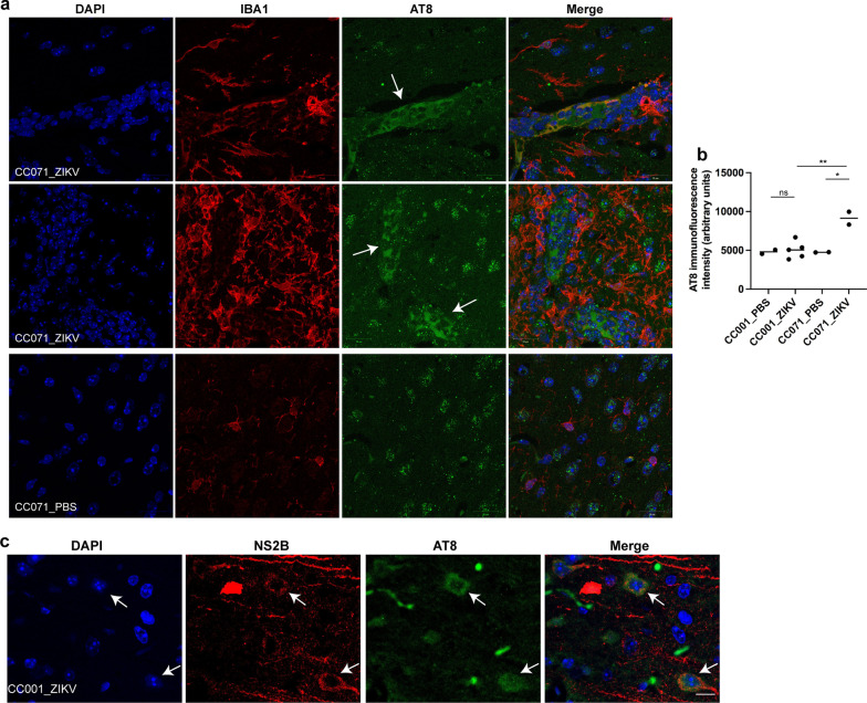
ZIKV infection induces pathological Tau phosphorylation in vivo. CC001 and CC071 mice (5–6 week-old) were necropsied at day 6 following IC inoculation of either PBS or 105 FFU of ZIKV. In vivo, ZIKV infection induced pTau recognized by AT8 antibodies predominantly in the brain of CC071 mice as determined by immunofluorescence and confocal microscopy with a, c nuclear DNA labeled with DAPI (blue), pTau labeled with the AT8 antibody (green), a microglial cells labeled with anti-Iba1 antibody (red) and c ZIKV-infected neurons labeled with anti-NS2B antibody (red). Arrows indicate in a clusters of pTau positive CC071 neurons surrounded by active microglial cells and in c ZIKV-infected pTau positive CC001 neurons. Single confocal sections (z projection) and the corresponding merge images are shown. Scale bars = 20 µm. b AT8 immunofluorescence intensity was quantified in cells from CC001 PBS-treated (n = 2 brains, 42 cells), CC001 ZIKV-infected (n = 5 brains, 107 cells), CC071 PBS-treated (n = 2 brains, 41 cells) and CC071 ZIKV-infected (n = 2 brains, 277 cells) mice. Symbols represent average values obtained for each brain with significance assessed by unpaired t-test. Means are indicated in graph. P-value < 0.01 (**), < 0.05 (*) and ns not significant

