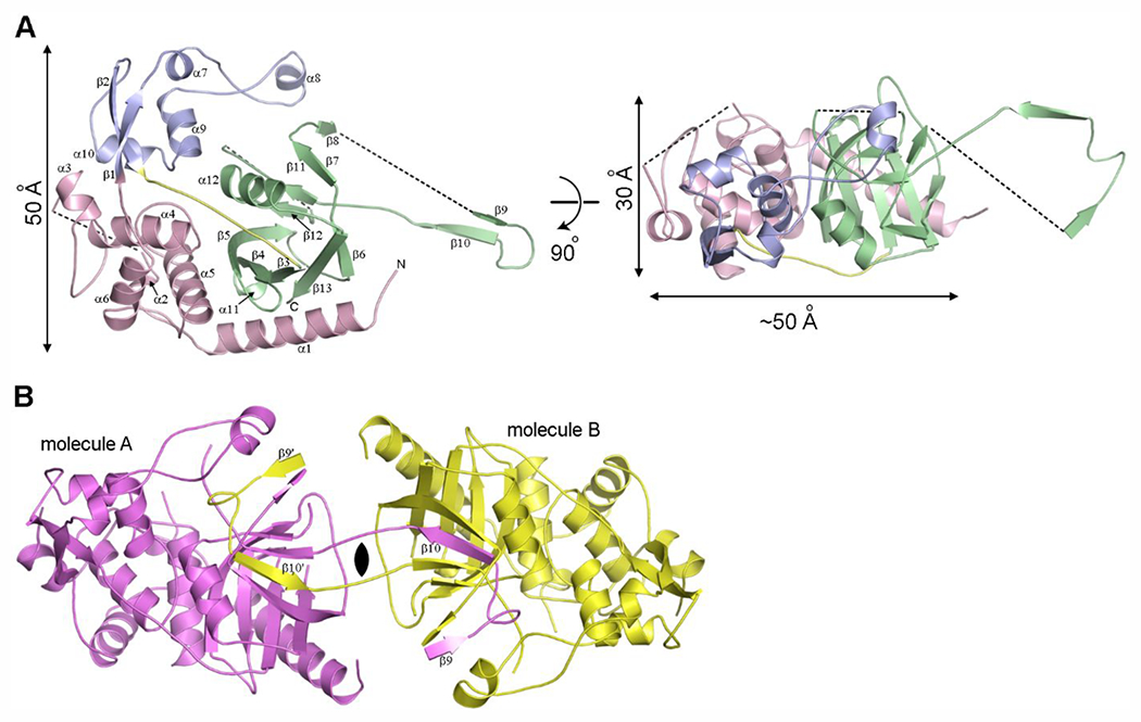
Figure 3.

The structure of LdtAb. (A) Ribbon representation of LdtAb colored by the structural domains; Domain-1 in pink, Domain-2 in blue, and Domain-3 (the catalytic domain) in green. The 10-residue linker connecting Domain-2 to the catalytic domain is shown in yellow. The black dashed lines indicate loops that are not visible in the electron density. The structure on the right is rotated approximately 90° relative to the structure on the left. (B) The crystallographic dimer generated from the LdtAb monomer (molecule A, magenta) showing the domain-swapped β9-loop-β10 motif from molecule A embedded in the symmetry-related molecule B (yellow).
