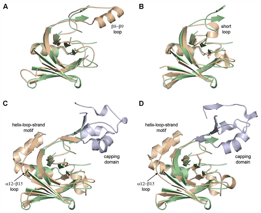
Figure 4.

The LDT catalytic domain. (A) Superposition of LdtAb (green ribbons) on K. pneumoniae YbiS (tan, PDB ID 4LZH). In the latter enzyme, the β8–β9 loop is a single α-helix. (B) Superposition of LdtAb (green ribbons) on B. subtilis YkuD (tan, PDB ID 1Y7M). (C) Superposition of LdtAb (green ribbons) on E. coli YcbB (tan, PDB ID 6NTW). (D) Superposition of LdtAb (green ribbons) on C. rodentium YcbB (tan, PDB ID 7KGM). In panels C and D the large capping domains are colored light blue, and the location of the second insertion is shown as the helix-loop-strand motif.
