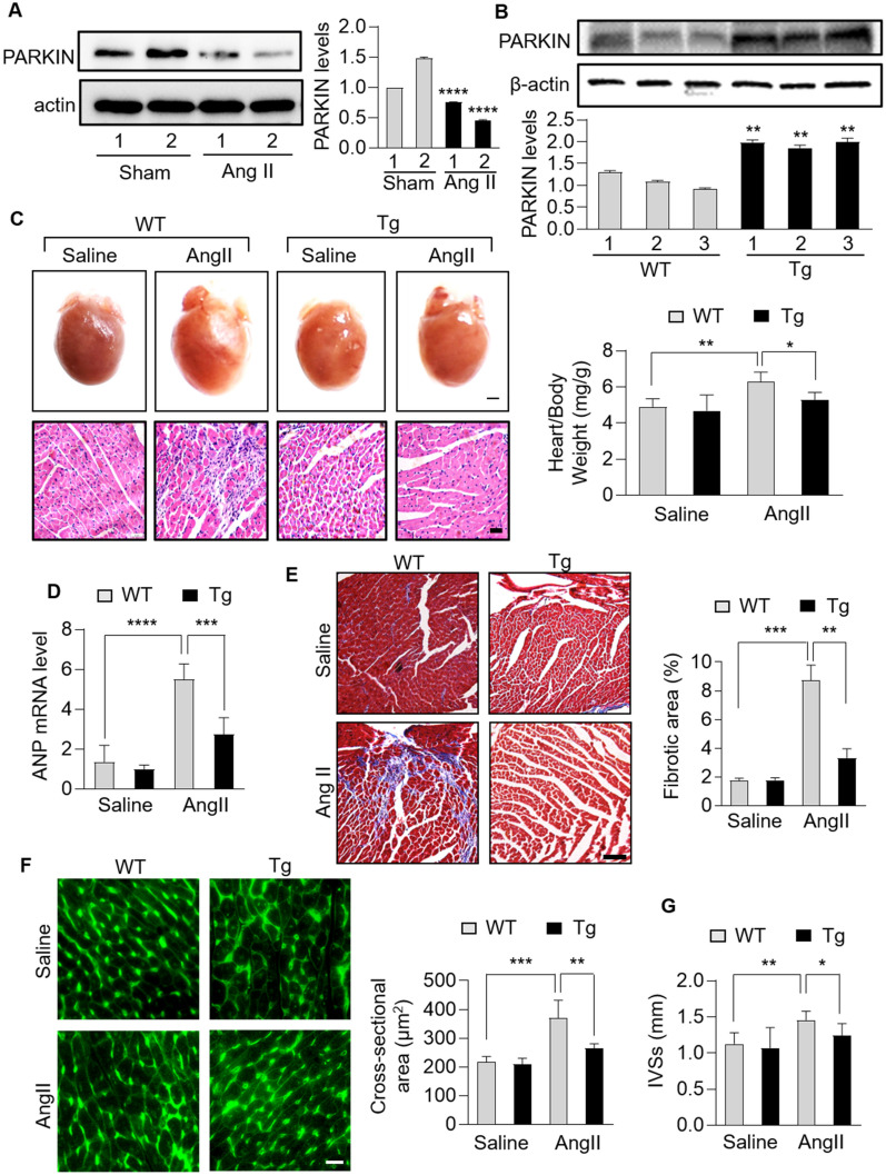
Fig. 1.
PARKIN negatively regulated cardiac hypertrophy in mice. A, Immunoblotting results showing the protein levels of PARKIN in mice hearts infused with angiotensin (Ang II) or not. n = 3 experiments per group. B, Immunoblotting results showing the protein levels of PARKIN in hearts of Parkin transgenic mice or wide-type (WT) mice. n = 3 experiments per group. C − E, Parkin transgenic mice exhibited reduced hypertrophic responses and cardiac fibrosis. Parkin transgenic mice and WT mice were infused with Ang II. C Top row: gross hearts (bar = 1.5 mm); bottom row: heart sections stained with hematoxylin and eosin (bar = 20 µm); right: the ratio of heart weight to body weight. * p < 0.05. ** p < 0.01. n = 5 experiments per group. D The mRNA levels of atrial natriuretic peptide (ANP) and brain natriuretic peptide (BNP) detected by qRT-PCR. The results were normalized to GAPDH. *** p < 0.001. ****p < 0.0001. n = 6 experiments per group. (E) Heart sections stained with Masson trichrome (left) and the fibrotic area analysis (right) (bar = 50 µm); ** p < 0.01. *** p < 0.001. n = 5 experiments per group. F and G, Parkin transgenic mice exhibited reduced cardiac remodeling and improved cardiac function in response to Ang II. F TRITC-conjugated wheat germ agglutinin staining was used to assessed cross-sectional area of hearts from WT mice and Parkin transgenic mice with Ang II infusions (left). The cross-sectional area was calculated (right). Bar = 100 µm; ** p < 0.01. *** p < 0.001. n = 6 experiments per group. G End-systolic interventricular septum thickness (IVSs) was measured. * p < 0.05. ** p < 0.01. n = 6 experiments per group

