Fig. 6.
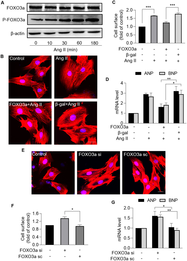
FOXO3a protected against cardiomyocyte hypertrophy. A, Immunoblotting results of the protein levels of total FOXO3a and phosphorylated FOXO3a in cardiomyocytes treated with Ang II at the indicated time. n = 3 experiments per group. B − D, Overexpression of FOXO3a inhibited Ang II-induced cardiomyocyte hypertrophy. Cardiomyocytes were exposed to Ang II after infected with FOXO3a adenovirus or β-gal adenovirus. B Sarcomere organization stained with phalloidin-TRITC conjugate; bar = 20 µm. Blue represent nucleus. Red represent F-actin. C Cell surface was calculated. *** p < 0.001. n = 3 experiments per group. D The mRNA levels of ANP and BNP were analyzed by qRT-PCR. The results were normalized to GAPDH. * p < 0.05. ** p < 0.01. n = 3 experiments per group. E − G, Knockdown of FOXO3a induced hypertrophic responses. Cardiomyocytes were infected with FOXO3a siRNA or FOXO3a scramble. E Sarcomere organization stained with phalloidin-TRITC conjugate; bar = 20 µm. Blue represent nucleus. Red represent F-actin. F Cell surface was calculated. * p < 0.05. n = 3 experiments per group. G The mRNA levels of ANP and BNP were analyzed by qRT-PCR. The results were normalized to GAPDH. * p < 0.05. ** p < 0.01. n = 3 experiments per group

