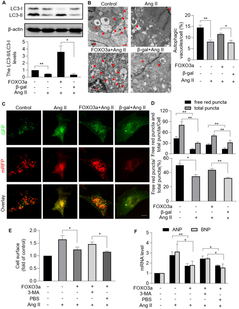
Fig. 7.
FOXO3a regulated cardiomyocyte mitophagy under hypertrophic stress. A, Enforced expression of FOXO3a rescued Ang II-induced decreased LC3II/LC3I. Cardiomyocytes were exposed to Ang II after infected with FOXO3a adenovirus or β-gal adenovirus. Immunoblotting was performed to detect protein levels of LC3I and LC3II (top). The ratio of LC3II/LC3I was calculated (bottom). * p < 0.05. ** p < 0.01. n = 3 experiments per group. B, FOXO3a restored autophagic vacuoles in hypertrophic model. Autophagic vacuoles were visualized in cardiomyocytes infected with FOXO3a adenovirus or β-gal (left; bar = 500 nm). Quantitation of autophagic vacuoles were shown in (right). * p < 0.05. ** p < 0.01. n = 3 experiments per group. C and D, Overexpression of FOXO3a attenuated Ang II-induced mitophagy flux defects. LC3 adenovirus tandem-labeled green fluorescent protein (GFP)-monomeric red fluorescent protein (mRFP) (GFP-mRFP-LC3) was used to indicate mitophagy flux. GFP-mRFP-LC3 was expressed and detected in 24 h after transfection in cardiomyocytes with overexpression or FOXO3a or not. GFP-LC3 (green puncta), mRFP (red puncta, representative of autolysosomes) and overlay (yellow puncta, representative of autophagosomes) (C; bar = 10 µm). Quantification results were shown in D. * p < 0.05. ** p < 0.01. n = 3 experiments per group. E and F, FOXO3a regulated cardiac hypertrophy through targeting mitophagy. After being infected with FOXO3a adenovirus, cardiomyocytes were treated with 3-Methyladenine (3-MA) or PBS, and then exposed to Ang II. (E) Cell surface area indicated by phalloidin-TRITC conjugate stained F-actin were calculated. *p < 0.05. n = 3 experiments per group. F The mRNA levels of ANP and BNP were detected. * p < 0.05. ** p < 0.01. n = 3 experiments per group

