Fig. 2.
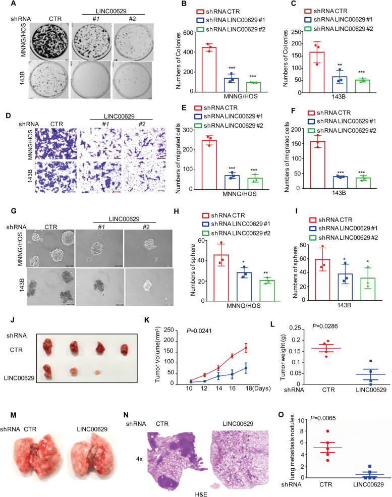
LINC00629 promotes osteosarcoma tumorigenesis and metastasis in vitro and in vivo (A-C) Osteosarcoma cells (3000 cells/well) with or without LINC00629 depletion were tested for cell growth in the colony formation assay. After 1 week, viable colonies were counted and are shown (A). Data are depicted as bar graphs (B-C). D-F The migration of the indicated cells was detected by Transwell assays. Representative images of crystal violet-stained culture plates are shown (D). Data are depicted as bar graphs (E, F). G-I The sphere formation abilities were detected in MNNG/HOS and 143B cells with or without LINC00629 knockdown. Representative images of the spheres are shown (G). Data are depicted as bar graphs (H-I). J-L MNNG/HOS cells (106 cells per mouse) with or without LINC00629 knockdown were injected subcutaneously into nude mice (n = 4). Representative images of xenograft tumours (J). The volume (K) and weight (L) of the tumours were calculated and analysed. M-O MNNG/HOS cells (106 cells per mouse) with or without LINC00629 knockdown were injected intravenously into nude mice (n = 5 per group). Representative images of lung (M) and HE (N) staining are displayed. Each group of metastatic nodules was assessed (O). Data in B, C, E, F, H, I, K, L and O were analysed by Student’s t test, *p < 0.05, **p < 0.01, ***p < 0.001

