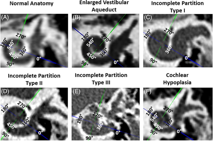
FIGURE 2.

Angular turn of the lateral‐wall traced manually with the help of cross‐hair feature (screenshots from OTOPLAN®) from different inner ear malformation types other than common cavity and vestibular cavity. Normal anatomy (A), enlarged vestibular aqueduct (B), incomplete partition type I (C), incomplete partition type II (D), incomplete partition type III (E), and Cochlear hypoplasia (F)
