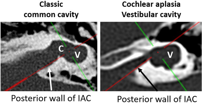
FIGURE 5.

Red line drawn along the posterior edge of the internal auditory canal in the axial view from OTOPLAN® distinguishes the cochlear (C) and the vestibular portion (V) in classic common cavity from the isolated vestibular portion in cochlear aplasia vestibular cavity. 13
