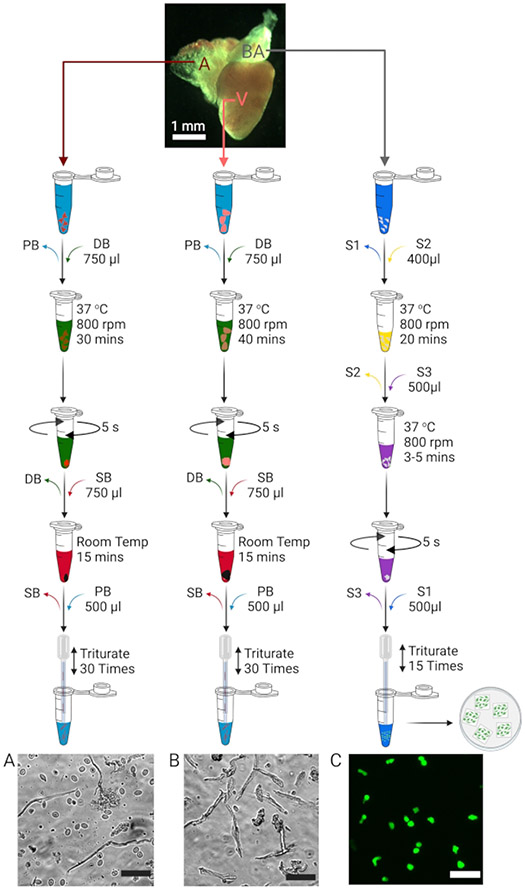
Figure 1: Isolation of atrial, ventricular, and bulbous arteriosus cells.
Schematic to illustrate the isolation of atrial (A), ventricular (B), and bulbous arteriosus (C) cells. The images depicting each isolated cell type (bottom) have scale bars = 50 μm in each case. BA cells isolated from smooth muscle cell transgenic reporter zebrafish lines (Tg(tagln:egfp)11, 12) are positive for green fluorescence.

