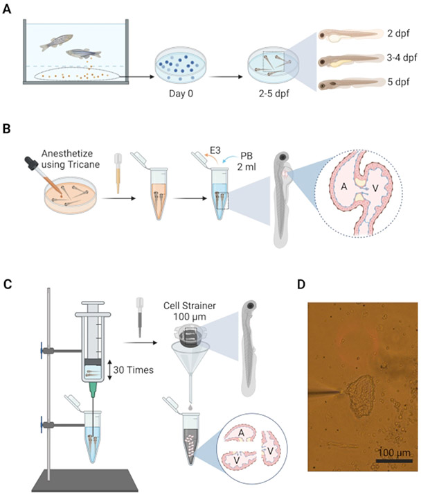
Figure 2: Schematic to illustrate the isolation of embryonic hearts.
(A) Fertilized embryos are removed from the breeding tank and placed in a Petri dish containing E3 water and maintained at 28 °C for up to 5 days. (B) At desired age, embryos are anesthetized in situ using tricaine and transferred into PB in 5 mL centrifuge tubes for dissociation. (C) Embryonic heart isolation apparatus consists of a 10 mL syringe attached to a 19 G needle mounted above the dissociation tube on a bench stand, allowing the hands to perform the trituration. (D) Image of the isolated heart (day 4) attached to a patch-clamp pipette.

