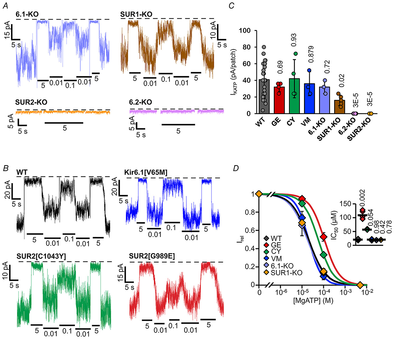
Figure 4. Subunit dependence of KATP channels in ventricular myocytes.

A and B, representative inside-out patch-clamp recordings from VCM in the presence of differing [ATP], as in Fig. 2A, from KATP subunit knockout fish (A) and Cantú mutant fish (B). with (right) or without (left) Mg2+ ions. C, measured KATP density (current in zero ATP) from individual experiments as in A and B (n = 3–24 recordings from 1–3 preparations (three animals per preparation), in each case). D, [ATP] dependence of channel activity (Irel) from recordings as in A,B. (above) Dose-response relationships were fitted with Hill plots (eqn (1)). Graph shows fit to averaged data; inset shows individual data, mean and SD for fits to individual recordings.
