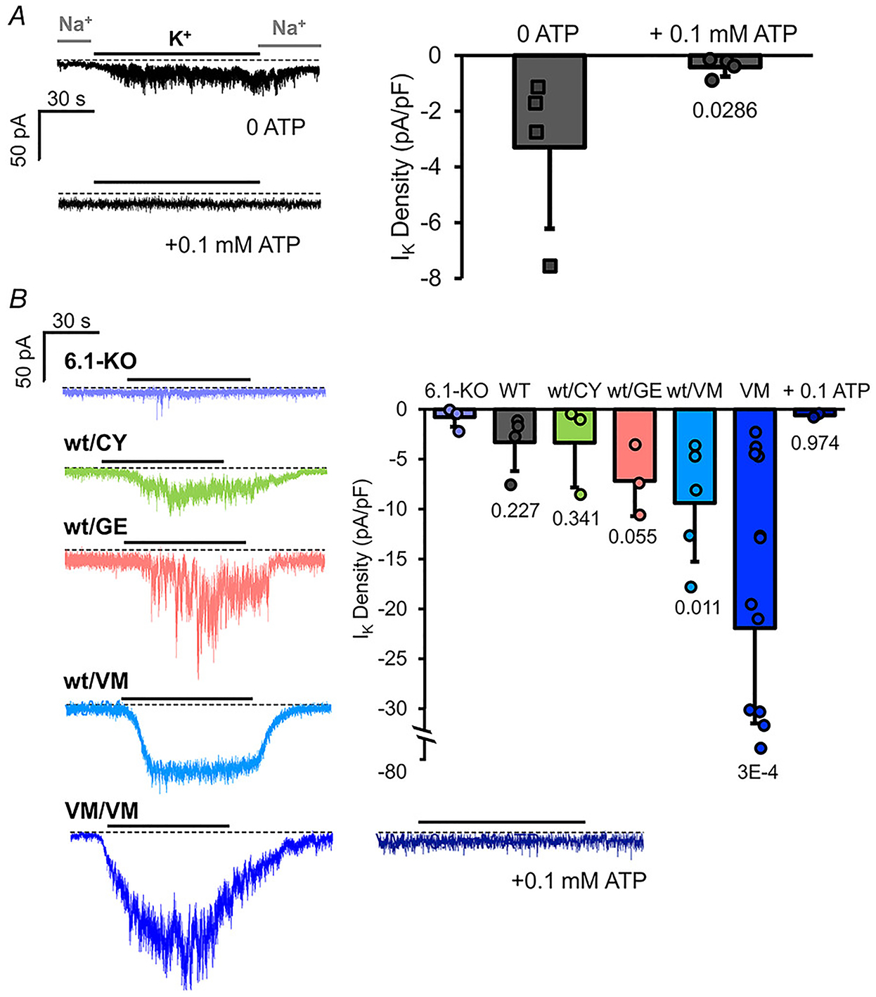
Figure 6. Subunit dependence of KATP channels in bulbous arteriosus myocytes.

A, left: representative whole-cell voltage-clamp recordings from WT BA smooth muscle myocytes with zero 0.1 mM ATP in the recording pipette. Currents were recorded with 136 mM Na+/6 mM K+ (Na+) in the bath or 0 Na+/140 K+ (K+) as indicated. Right: measured K current density (current in K+ – current in Na+) from individual experiments as at left (n = 4 recordings from 2 preparations (five animals per preparation), in each case). B, left: representative whole-cell voltage-clamp recordings from Kir6.1 knockout and hetero- or homozygous Cantú mutant zebrafish as indicated. Right: measured K+ current density (current in K+ – current in Na+) from individual experiments as at left (n = 3–10 recordings from 1–3 preparations (five animals per preparation), in each case).
