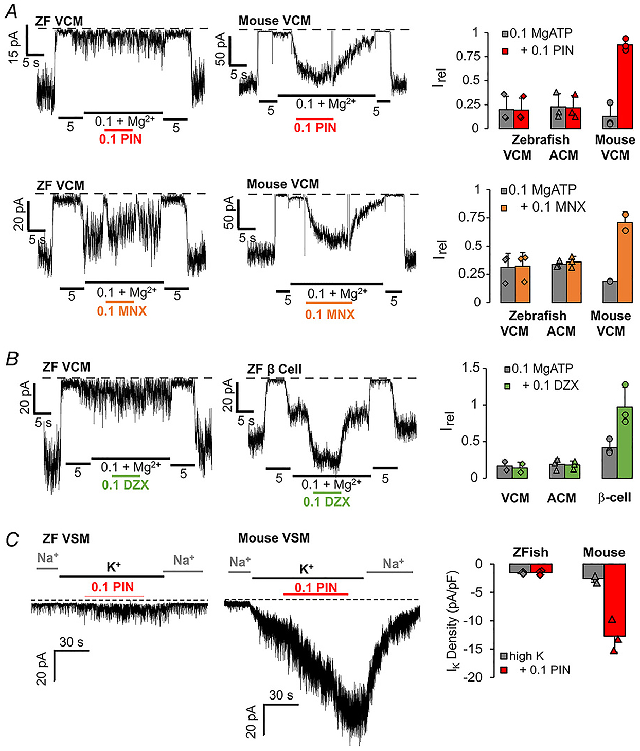
Figure 7. K+ channel opener insensitivity in zebrafish cardiovascular myocytes.

A, left: representative inside-out patch-clamp recordings from zebrafish and mouse VCM, as indicated, in the presence of differing [ATP], with or without pinacidil (0.1 mM, PIN) or minoxidil (0.1 mM MNX). Right: currents in 0.1 mM ATP with and without drug (Irel). Graph shows individual data, mean and SD (n = 3, in each case). B, left: representative inside-out patch-clamp recordings from zebrafish VCM and pancreatic β-cell, as indicated, in the presence of differing [ATP], with or without diazoxide (0.1 mM, DZX). Right: currents in 0.1 mM ATP with and without DZX (Irel). Graph shows individual data, mean and SD (n = 3 recordings from 1 preparation each), in each case). C, left: representative whole-cell voltage-clamp recordings from zebrafish BA (VSM) and mouse aortic VSM, as indicated, with or without addition of pinacidil (0.1 mM, PIN). Right: K+ current density in high K+ solution, with and without PIN. Graph shows individual data, mean and SD (n = 3 recordings from 1 preparation in each case).
