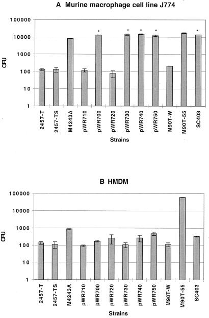
FIG. 1.
Evaluation of infection of ipaH mutant strains in J774 cells (A) and HMDM (B). Bacteria were left in contact with macrophages for 30 min, washed with HBSS, and further incubated in gentamicin-containing medium for another 50 min. Macrophages were then washed and lysed. The numbers of viable bacteria were obtained by plating dilutions of the lysates on TSA plates. CFU represents the total number of bacteria in macrophage cell lysates. The characteristics of the strains are listed in Table 1. ∗, P value not significant compared to M90T-55. Error bars show means ± standard deviations.

