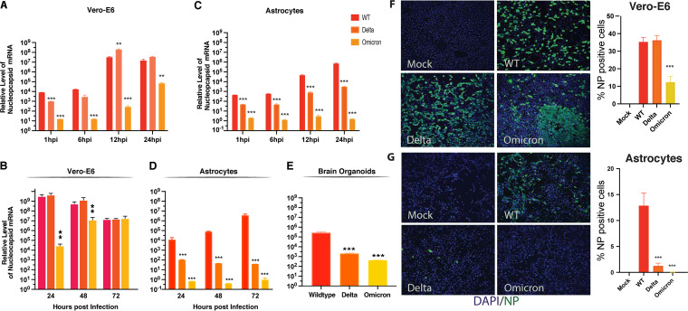
FIG 3.
Delta and Omicron variants of concern display less efficient replication in astrocytes and brain organoids. (A) Vero-E6 cells were infected with the indicated viruses at a multiplicity of infection (MOI) of 0.01. Cell lysates were collected 1, 6, 12, and 24 h after infection. The expression of nucleocapsid viral RNA was measured by RT-qPCR and normalized based on the mock infection control. *, P < 0.05; **, P < 0.01; ***, P < 0.001 (by Student’s two-tailed t test). (B) Vero-E6 cells were infected with the indicated viruses at an MOI of 0.01. Cell lysates were collected 24, 48, and 72 h after infection. The expression of nucleocapsid viral RNA was measured by RT-qPCR and normalized to the mock infection control. *, P < 0.05; **, P < 0.01; ***, P < 0.001 (by Student’s two-tailed t test). (C) Astrocytes were infected with the indicated viruses at an MOI of 0.01. Cell lysates were collected at 1, 6, 12, and 24 h postinfection. The expression of nucleocapsid viral RNA was measured by RT-qPCR and normalized to the mock infection control. *, P < 0.05; **, P < 0.01; ***, P < 0.001 (by Student’s two-tailed t test). (D) Astrocytes were infected with the indicated viruses at an MOI of 0.01. Cell lysates were collected at 24, 48, and 72 dpi. The expression of nucleocapsid viral RNA was measured by RT-qPCR and normalized to the mock infection control. *, P < 0.05; **, P < 0.01; ***, P < 0.001 (by Student’s two-tailed t test). (E) four-month-old brain organoids were infected with the indicated viruses at an MOI of 1. After 6 h of incubation, the medium was replaced. Cell lysates were collected at 7 days postinfection. The expression of nucleocapsid viral RNA was measured by RT-qPCR and normalized to the mock infection control. *, P < 0.05; **, P < 0.01; ***, P < 0.001 (by Student’s two-tailed t test). (F) Vero-E6 cells were infected with the indicated viruses at an MOI of 0.1. Cells were fixed and immunostained at 48 h postinfection. Four image fields per group from two independent experiments were quantitated for the presence of NP. *, P < 0.05; **, P < 0.01; ***, P < 0.001 (by Student’s two-tailed t test). (G) Astrocytes were infected with the indicated viruses at an MOI of 0.1. Cells were fixed and immunostained at 48 h postinfection. Four image fields per group from two independent experiments were quantitated for the presence of NP. *, P < 0.05; **, P < 0.01; ***, P < 0.001 (by Student’s two-tailed t test).

