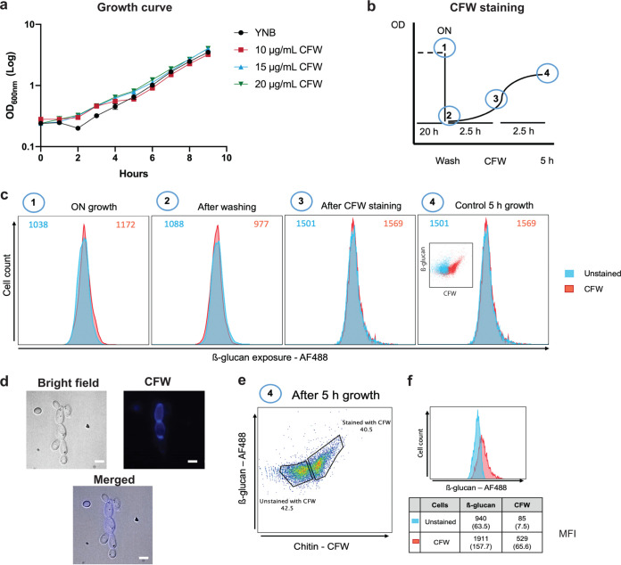
FIG 3.
Most glucan exposure is associated with mother cells. Calcofluor white (CFW) staining was used to differentiate older mother cells from younger daughter cells. (a) First, the effect of CFW on growth was tested. C. albicans SN250 cells were grown on GYNB after staining for 5 min with no CFW (yeast nitrogen base [YNB]) or with 10, 15, or 20 μg/mL CFW and washed, and then the growth in medium without CFW was monitored for 9 h. (b) Scheme showing the experimental design. C. albicans SN250 cells were grown overnight in GYNB (1) and then washed and transferred to fresh GYNB at an OD600 of 0.2 (2). The cells were then grown for 2.5 h and stained with 20 μg/mL CFW for 5 min (3). The cells were then grown for a further 2.5 h (approximately one doubling time) (4). (c) A control experiment was performed to test whether CFW staining in the context of this experimental design affects subsequent staining with Fc-dectin-1. At each stage (labeled 1 to 4), cultures were split in two: one part was stained for 5 min with 20 μg/mL CFW, whereas the other part was not. Each sample was grown as indicated in GYNB, stained with Fc-dectin-1, and analyzed by flow cytometry. (d) Representative images from microscopy showing C. albicans SN250 wild-type strain presenting yeast and budding yeast forms in bright-field, CFW-stained (mother cells), and merged images (scale bar, 5 μm). (e) After performance of these control experiments, experiments were performed as described for panel c. Then, C. albicans SN250 cells were gated based on their CFW staining, and the levels of β-1,3-glucan exposure were determined for the CFW-positive and CFW-negative subpopulations. (f) The mean CFW MFI and Fc-dectin-1 MFI (with standard deviation) from three independent experiments are shown for each subpopulation.

