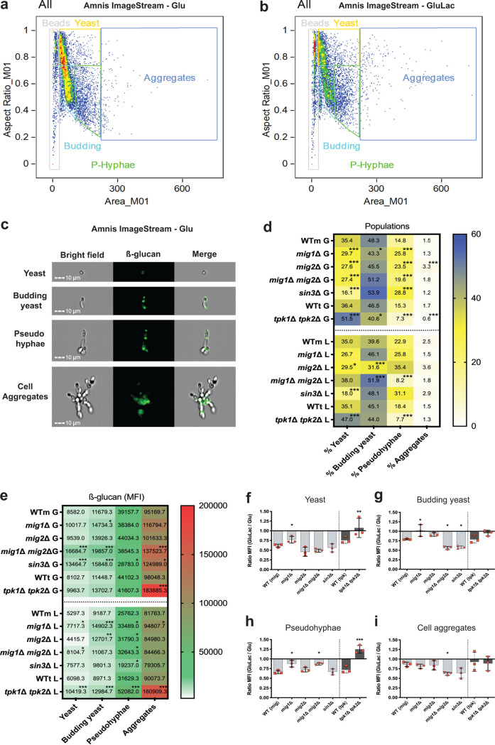
FIG 8.
Regulation of β-1,3-glucan exposure in yeast and pseudohyphal cells. C. albicans cells were grown for 5 h in GYNB or GYNB containing lactate and stained with Fc-dectin-1. The proportions of yeast and pseudohyphal cells in these populations, and their levels of β-1,3-glucan exposure, were then analyzed by imaging flow cytometry. Based on cell morphology observed by imaging, and the distribution of the cells by cytometry, cell populations were gated into unbudded yeast cells, budding yeast, pseudohyphae, and filamentous cells. (a) Gating of cells grown on GYNB (glucose alone). (b) Gating of cells grown on GYNB containing lactate. (c) Representative images of C. albicans SN250 cells in these subpopulations, including bright-field, fluorescent micrograph, and merged images. (d) Percentages of unbudded yeast cells, budding yeast cells, pseudohyphae, and filamentous cells observed in different C. albicans strains grown for 5 h on GYNB (G) or GYNB containing lactate (L), including WTm (SN250), mig1Δ, mig2Δ, mig1Δ mig2Δ, sin3Δ, WTt (SN152HLA), and tpk1Δ tpk2Δ strains. (e) Levels of β-1,3-glucan exposure (MFI) displayed by each subpopulation shown in panel d. (f to i) Fold change in β-1,3-glucan exposure for each subpopulation shown in panel e, calculated as the ratio of GluLac MFI to Glu MFI. Means and standard deviations of results from three independent experiments are shown. The data in panels d to f represent means of results from three independent experiments and were analyzed using two-way ANOVA with Tukey’s multiple-comparison test (*, P < 0.05; **, P < 0.01; ***, P < 0.001).

