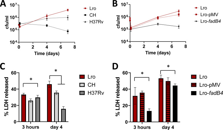
FIG 3.
Lro increased more rapidly than did the comparator strains in THP-1 monocytes and caused greater cell damage. The THP-1 were primed with 10 ng/mL INF-γ and were then infected with the indicated strains of M. tuberculosis at a 1:1 ratio. (A and B) Intracellular bacteria were recovered via the lysis of THP-1 and quantitated by CFU. Data are the means and standard deviations of at least four infections. At 7 days, the CFU values for Lro were significantly higher than those for CH, H37Rv, or Lro-fadB4 (P < 0.05). (C and D) Cytotoxicity was determined via the measurement of lactate dehydrogenase activity in the cell culture medium of infected cells and was compared to the LDH activity when 100% of the cells were lysed using detergent. 100% represents the LDH activity when THP-1 were fully lysed using detergent. Data are the means and standard deviations of at least four wells. An asterisk indicates P < 0.05.

