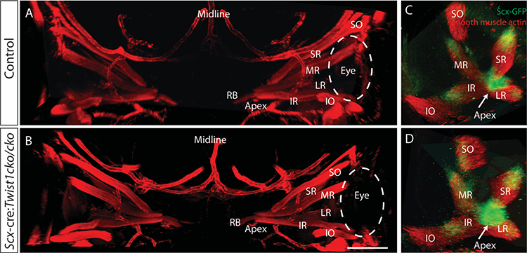
Figure 4.
Loss of Twist1 in Scx expressing cells does not disrupt EOM organization or tendon formation. Thick sections from E14.5 embryos stained with anti-smooth muscle actin (red) and imaged coronally show the normal arrangement of the EOMs in both control embryos (A) and Scx-cre:Twist1cKO/cKO embryos (B). Orbits from whole mounts of E13.5 embryos expressing GFP from the scleraxis promoter show normal tendon formation at the apex (arrow) and end of each muscle in both control embryos (C) and Scx-cre:Twist1cKO/cKO embryos (D). Images in C and D were reconstructed in Arivis and are oriented looking into the orbit towards the apex. Abbreviations as per Figure 1. n= 5 Scx-cre+, 3 littermate controls. Scale bar in B equals 100um.

