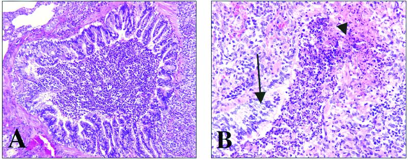
FIG. 2.
Representative photomicrographs of a lung from a guinea pig vaccinated with the hsp60-hsp70 protein mixture. (A) Terminal bronchiole surrounded by granulomatous-lymphocytic inflammation. The lumen is filled with mucus, sloughed epithelium, and necrosuppurative cellular debris. Magnification, ×160. (B) Terminal bronchiole with focal epithelial ulceration (arrowhead) and focal epithelial hyperplasia (arrow). The lumen is again filled with mucus, sloughed epithelium, and necrosuppurative cellular debris. Magnification, ×320. Hematoxylin and eosin staining was used.

