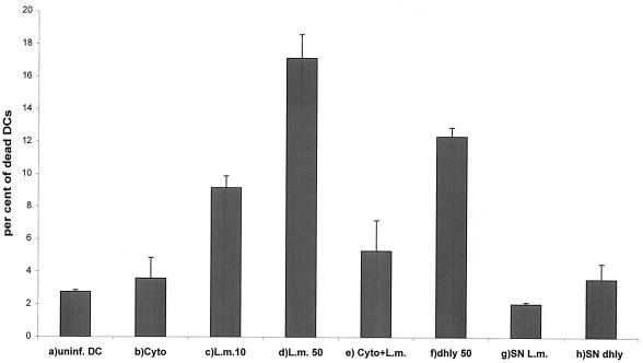
FIG. 8.
Determination of the percentage of dead DCs at 6 h p.i. by flow cytometry after propidium iodide staining. (a) Uninfected DCs (negative control). (b) Uninfected DCs after treatment with cytochalasin D (2 μg/ml). (c) L. monocytogenes wild type (10403S) infection at an MOI of 10. (d) L. monocytogenes wild type (10403S) infection at an MOI of 50. (e) L. monocytogenes wild type (10403S) infection at an MOI of 50 after pretreatment with cytochalasin D (2 μg/ml). (f) L. monocytogenes Δhly infection at an MOI of 50. (g) Addition of L. monocytogenes wild type supernatant (20%). (h) Addition of L. monocytogenes Δhly supernatant (20%). Results are presented as the mean values and the standard deviation (error bars) of three independent experiments.

