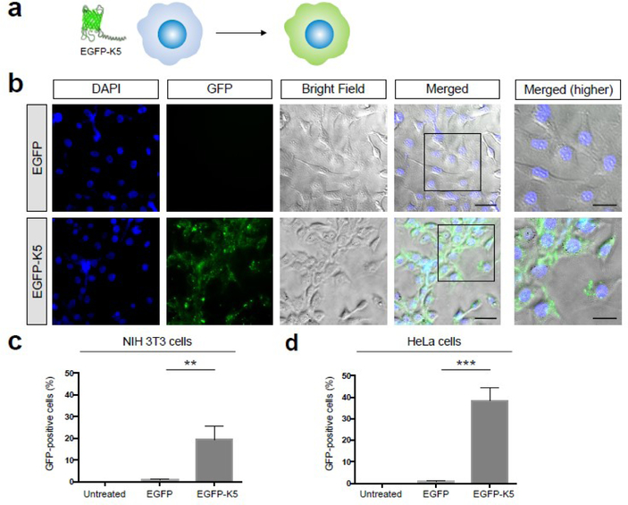
Figure 2. K5 enhances the delivery of EGFP into various cell types.
(a) Schematic illustration of enhanced cellular uptake of EGFP by K5. (b) K5 facilitates the internalization of EGFP into NIH 3T3 cells. Higher magnifications of the merged images (black boxes) are shown in the right panel. Scale bars represent 50 μm (left panels) and 30 μm (right panels), respectively. (c-d) Quantification of flow cytometry data of EGFP or EGFP-K5 uptake in (c) NIH 3T3 cells and (d) HeLa cells. Mean ± SEM. ** P < 0.01, *** P < 0.001 by permutation t-test. P values were calculated between EGFP and EGFP-K5.

