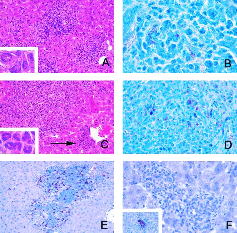
FIG. 2.
Granulomatous lesions in GKO mice infected with M. genavense. GKO mice were infected with 106 M. genavense cells, and livers and lymph nodes were removed for histological analysis at indicated times postinfection. (A) Malorganized inflammatory infiltrations in the liver composed predominantly of mononuclear cells at 20 weeks postinfection (hematoxylin and eosin stain; magnification, ×64). Inset, enlargement of intralesional mononuclear cells with little epithelioid transformation (magnification, ×320). (B) Granuloma in the liver with acid-fast M. genavense at 20 weeks postinfection (Ziehl-Neelsen stain; magnification, ×128). (C) Large diffuse mixed infiltrations and perivascular accumulation of predominantly polymorphonuclear cells (arrow) at 26 weeks postinfection (hematoxylin and eosin stain; magnification, ×64). Inset, enlargement of lesion indicated by arrow, demonstrating typical polymorphonuclear morphology (magnification, ×320). (D) Acid-fast M. genavense in mesenterial lymph node at 26 weeks postinfection (Ziehl-Neelsen stain; magnification, ×128). (E) CD3-positive cells in granulomatous lesions at 20 weeks postinfection (immunoperoxidase; magnification, ×64). (F) Granulomatous lesions negative for material reactive with an anti-iNOS-antiserum at 20 weeks postinfection (immunoperoxidase; magnification, ×128). Inset, iNOS-positive control in M. tuberculosis-infected BALB/c mice stained in parallel.

