Figure S1.
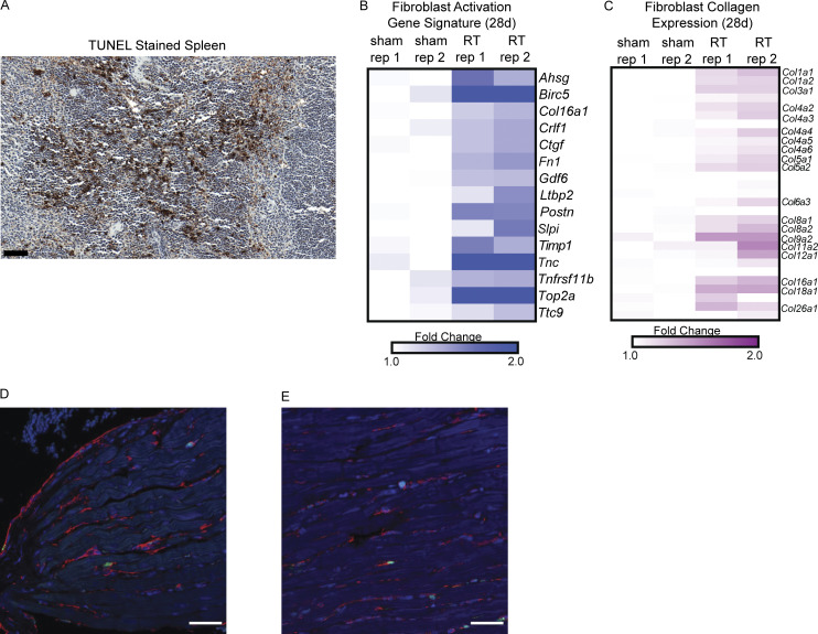
A mouse model of radiation-induced cardiac toxicity. (A) TUNEL staining in mouse spleen as positive control for cellular apoptosis in tissue. Scale bar = 50 μm. (B) Expression of genes associated with fibroblast activation in cardiac fibroblasts from C57BL/6 as defined in Park. et al. (2018) in fibroblasts isolated from hearts 28 d after sham treatment or RT. (C) Expression of collagen genes in fibroblasts isolated from hearts 28 d after sham treatment or RT. (D and E) Low magnification (40×) immunofluorescence images of hearts sections of Sting+/+ mice 28 d after sham treatment (D) and cardiac RT (E). cGAS, green; Vimentin, red; DAPI, blue. Experiment performed with N = 4 mice for each treatment condition and timepoint; representative image shown.

