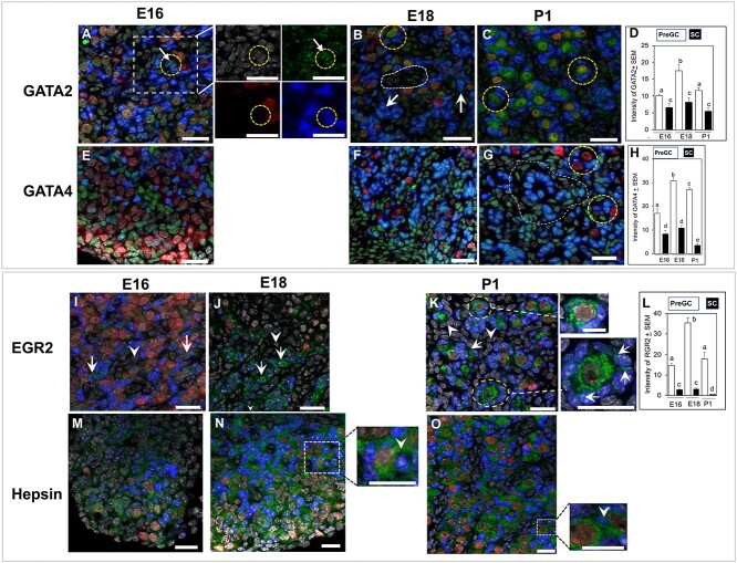
Figure 6.
GATA2, GATA4, EGR2, and hepsin localization in sections of E16, E18, and P1 ovaries. (A–D) Nuclear localization (circle) of GATA2 (green) protein in E16 (A), E18 (B), and P1 (C) ovaries and quantitation of its immunosignal intensity (D). Arrows show FOXL2-positive pre-GC in E16 ovary (A). Arrows showing GATA2 signal in FOXL2-positive pre-GC in an E18 ovary (B). Broken circles indicate GATA2 staining in the oocytes on E18 (B) and P1 (C) ovaries. Broken line encircles a pool of non-GC cells (B). High intensity of FOXL2 (blue) often masked green fluorescence of GATA2. (E–H) GATA4 (green) localization in E16 (E), E18 (F), and P1 (G) and quantitation of its immunosignal intensity (H). Intense ovarian GATA4 immunosignal was present in pre-GC of E18 (F) and P1 (G) ovaries. Broken circles (G) showed pre-GC associated with PFs. Higher intensity of FOXL2 immunosignal masked GATA4 immunosignal forming teal-colored nuclei and presented an apparent higher expression in non-GC (area encircled by dotted line; see Supplemental Figures S3 and S4 for clarification). However, the difference in GATA4 immunosignal between two cell types was prominent in the bar graph (H). (J–M) EGR2 localization in E16 (J), E18 (K), and P1 (L) ovaries and quantitation of its immunosignal intensity (L). Nuclear localization of EGR2 protein (green) in FOXL2-positive pre-GC (arrows) and non-GC (arrowheads) in the E16 mouse ovary (I). EGR2 immunosignal in pre-GC on E18 (J) and P1 (K) ovaries (arrows). Cytoplasmic immunosignal in P1 ovary (inset). Higher magnification images of primordial or primary follicles in the P1 ovary (K) (insets). (M-O) Hepsin (green) localization in sections of E16 (M), E18 (N), and P1 (O) ovaries. Inset showing higher magnification image of hepsin staining between adjacent pre-GC in E18 and P1 ovaries (inset, arrowhead). Bars with same letter, P > 0.05; bars with different letters, P < 0.05. Green, target proteins; blue, FOXL2; red, GCNA; gray, nuclei. Bar = 25 μm.

