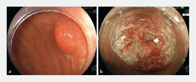
Fig. 1.

Images from magnifying narrow-band imaging (NBI) of a cecal lesion showing: a a 7-mm sessile tumor without a demarcated depressed area, which initially resembled a sessile serrated lesion and was suspected to be an adenocarcinoma on a previous biopsy; b under blue light, a vascular pattern with both interruption of thick vessels and loose vascular areas, and a surface pattern with amorphous areas, which was classified as type 3 (the Japan NBI Expert Team classification), consistent with a diagnosis of deep submucosal invasive cancer.
