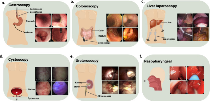
Fig. 2. Different endoscopic acquisition systems for various hollow organs.
a Gastroscopy procedure during which a flexible endoscope is inserted to visualise mucosa in the oesophagus and stomach parts of the duodenum. It can be observed that the scene varies quite a lot depending on the scope location. Similarly, in the top left image, one can observe bubbles surrounding the mucosa. b Colonoscopy procedures cover the colon and rectum, during which flexible endoscopes are used to navigate this complex twisted organ. Bowel cleansing is an essential preparation as it can occlude lesions. In most images, the presence of stool is a clear mark of occluded anomaly. c During laparoscopy, usually rigid endoscopes are inserted through small incision holes. Images depicting fat surrounding the liver, a clear view of the liver, the presence of tools during surgery and complete occlusion of the liver due to fat are shown. d Widely used rigid endoscopes are used for investigating bladder walls that are inserted through the urethra. Conventional white light image modality (first three) and fluorescence image (blue) modality are shown125. It can be observed that the top two images are blurry showing little or no vessel structures. e Kidney stone removal using ureteroscopy and laser lithotripsy. The difference in texture and surrounding debris (in top) and blood (bottom) for in vivo images71. f A flexible endoscope enters through the nostrils and can go from the nose up to the throat area and is hence collectively called nasopharyngeal endoscopy. Images (on the left) show a small opening and field of view, along with surgical tools for some cases126. The sources of relevant endoscopy images: gastroscopy and colonoscopy images in (a and b are acquired from Oxford University Hospitals under Ref. 16/YH/0247 and forms part of publicly released endoscopy challenge datasets (EDD2020127 under CC-by-NC 4.0 and PolypGen128 under CC-by, Dr S. Ali is the creator of both datasets). Liver laparoscopy data are taken from the recently conducted P2ILF challenge129 (Dr S. Ali is the creator of this dataset), while cystoscopy and ureteroscopy data are respectively taken from PhD thesis of Dr S. Ali130 and a recently published paper of which he is a co-author71. Similarly, nasopharyngeal images correspond to publicly available UW-Sinus-Surgery-C/L dataset126 with an unknown licence.

